Skip to content

CT of the head, nose and ears

A head CT shows the nasal cavity, middle ear, jaw with tooth roots, skull and brain. Bone and soft tissue in one examination, exactly where an X-ray is not enough. We scan dogs and cats under general anaesthesia, and a radiologist writes the report.

From €159 including anaesthesia, the radiologist's report and VAT

What the CT shows

Head, nose and ears

The nasal cavity in cases of nasal discharge or nosebleeds, the middle ear, the jaw and tooth roots, the skull and the brain. Bone and soft tissue in one examination, exactly where an X-ray is not enough.

Neck and larynx

Larynx, thyroid gland, pharynx, lymph nodes and foreign bodies.

Teeth and jaw

Teeth, roots and periodontium, jaw bones, where intraoral X-rays are not enough.

When a vet recommends a head CT

  • Nasal discharge or bleeding that does not go away
  • Middle ear infection that keeps coming back
  • Teeth and jaw when an intraoral X-ray is not enough
  • Neurological signs where the skull and brain need to be seen

Examination prices

Prices include anaesthesia and VAT. The lower price applies to a standard report, the higher one to a report within 24 hours. A patient over 8 years old or unwell needs a pre-anaesthetic examination before CT. We can do it here for €89, or you can upload results no older than 7 days. You will see the total in the order summary and pay it at the clinic at the examination. Details, including cancellation, are in the terms and conditions.

Real cases from our CT

Oncology, Abdomen, Head and neck

She was losing weight and vomiting blood. CT showed the problem was not just the thyroid

Patient
cat, domestic shorthair, spayed female, 16 years
Examination
Whole-body CT, pre- and post-contrast
Date
March 2026
3D CT reconstruction

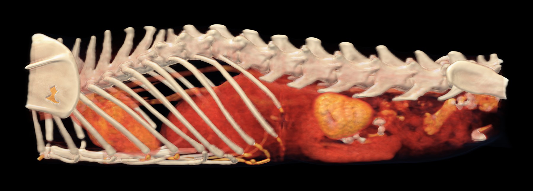
3D reconstruction of the chest and abdomen, seen from the left. Behind the ribs is the enlarged left kidney (gold), in which the CT found nodules; further forward, in the chest, is the heart.

  • bone
  • strongest contrast uptake: blood vessels, heart, kidneys
  • organs and tissues with weaker contrast uptake, including tumours
  • skin and fat

A sixteen-year-old cat was being treated for an overactive thyroid gland. Despite this, she was losing a lot of weight and had been vomiting blood for a long time. The vet needed to know what was happening throughout her body and therefore recommended a CT scan from head to tail.

In the neck, the CT found an enlarged right lobe of the thyroid gland, a mass about four centimetres long that was not growing into the surrounding tissue. However, it also showed things that an examination of the thyroid alone would not have revealed: a thickened stomach wall, enlarged lymph nodes in several places, lesions in the spleen, tiny nodules in the liver, nodules in the left kidney and a little fluid in the abdomen. The right kidney was small, which is consistent with a long-standing disease.
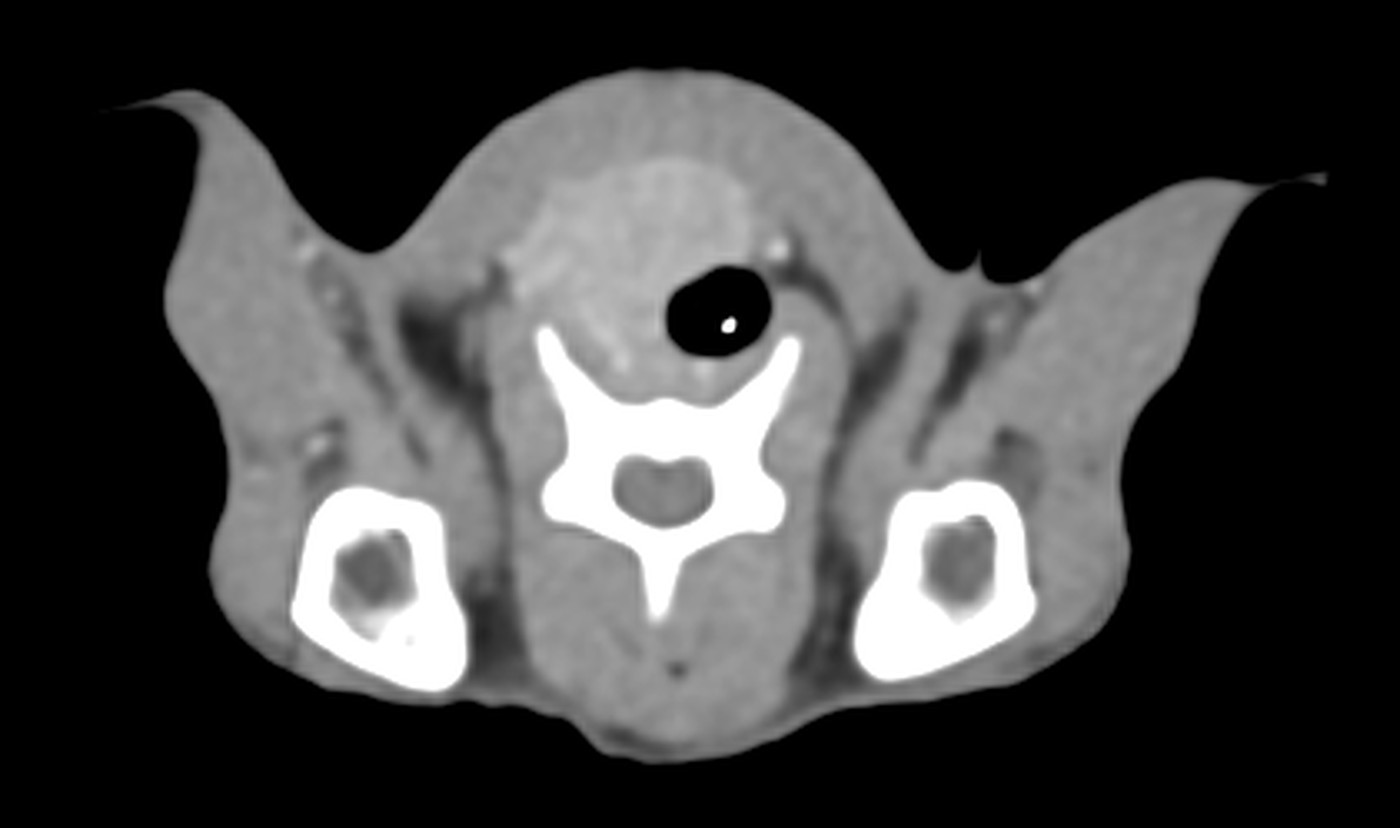
Transverse CT slice at the level of the finding

Cross-section through the neck: next to the windpipe is a non-uniform mass, the enlarged right lobe of the thyroid gland.
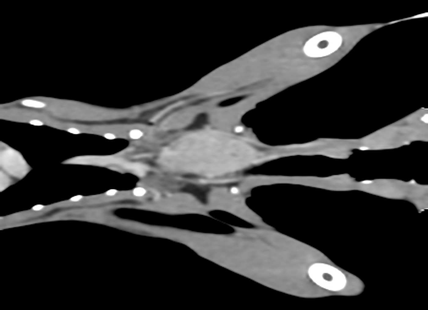
Longitudinal CT slice through the neck

Longitudinal slice through the neck, seen from above: the oval mass in the centre is the enlarged right lobe of the thyroid gland.
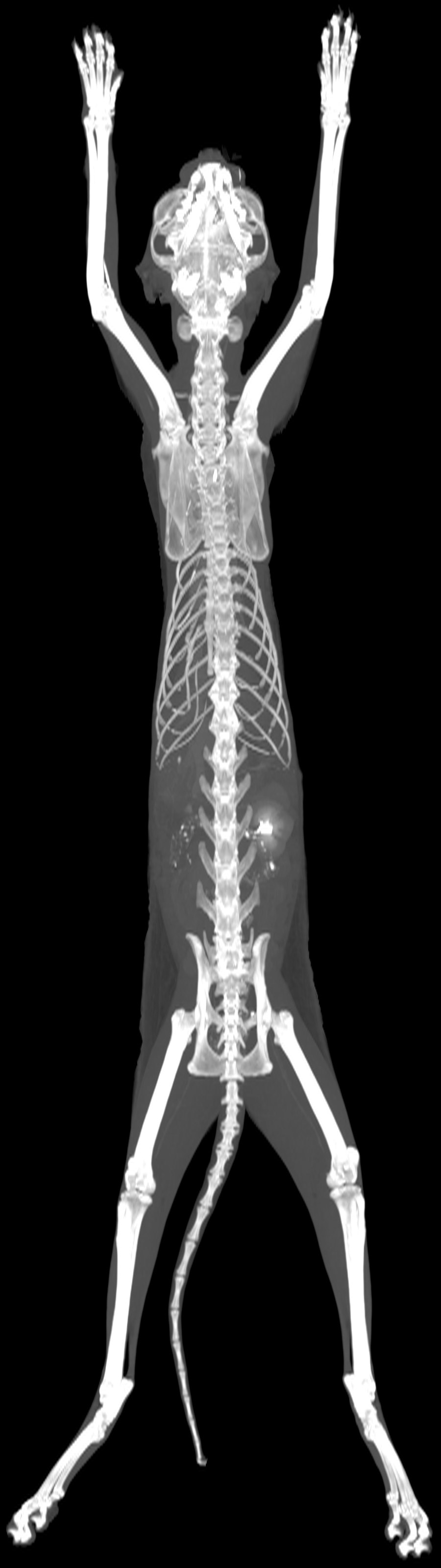
Maximum intensity projection of the whole scan

The whole scan in a single image: the skeleton, and the kidneys highlighted by the contrast agent.
Nose and ear, Chest and lungs, Oncology

He came in because of his ear. CT also found a mass near the heart

Patient
dog, French Bulldog, male, 7 years
Examination
Whole-body CT, pre- and post-contrast
Date
March 2026

A seven-year-old French Bulldog had started tilting his head to the right and had a discharge from his right ear. To find out what was going on in the ear and deeper behind it, he had a whole-body CT scan.

The CT confirmed inflammation of both outer ear canals, with a build-up of secretions on the right, and changes in the middle ear cavities that are common in flat-faced breeds. At the back of the nose it showed aberrant nasal turbinates, in other words tissue that should not normally be there.

The most important finding, however, was one the dog had not come in for: a mass about five centimetres in size at the base of the heart, wrapping around the aorta and pressing on both pulmonary arteries, and next to it fluid in the sac around the heart. An examination focused only on the ear would have left it hidden.

Head and neck, Chest and lungs, Abdomen

An itchy lump on the neck, and everything the CT showed

Patient
cat, British Shorthair, neutered male, 11 years
Examination
Whole-body CT, pre- and post-contrast
Date
March 2026
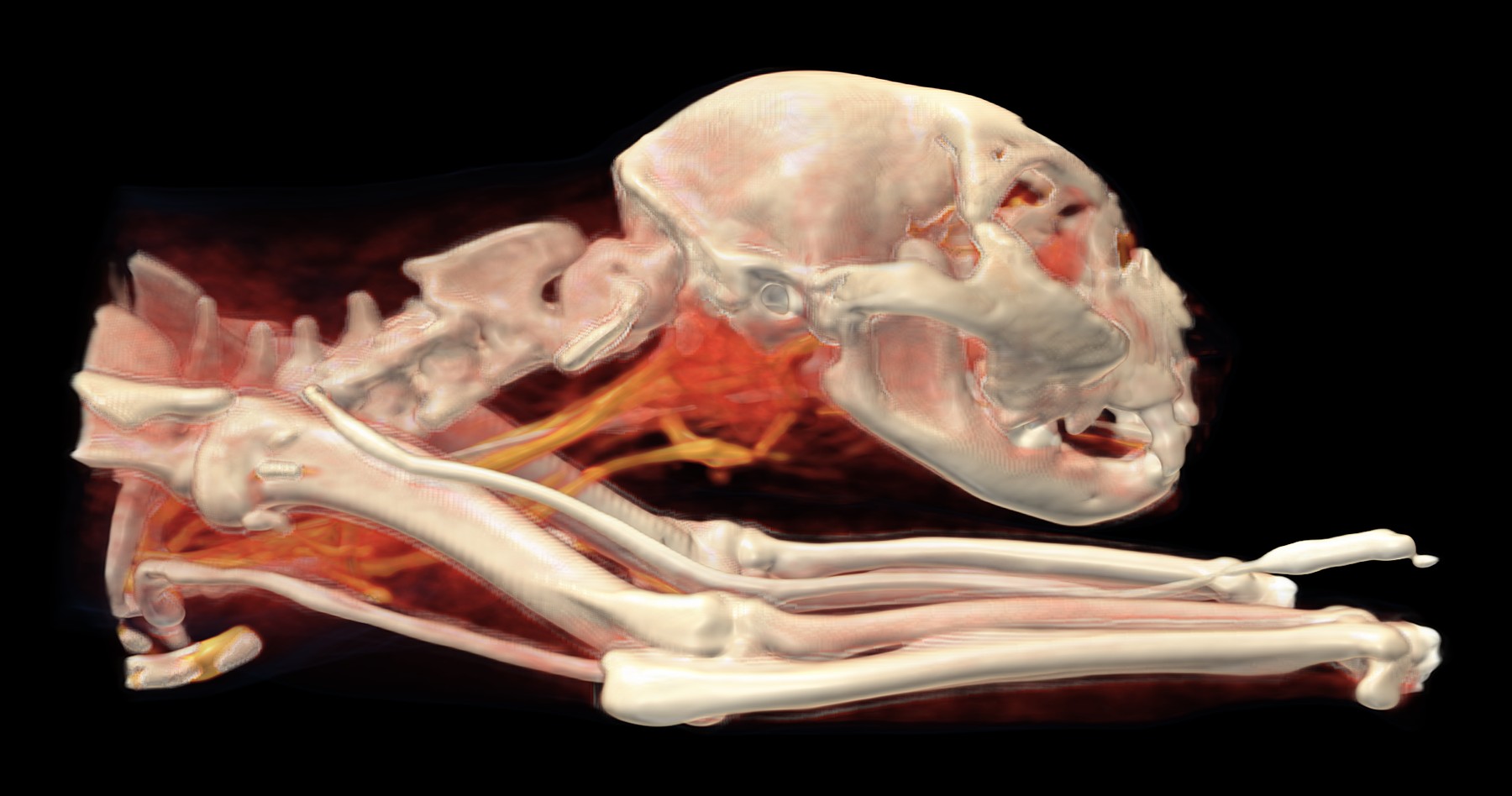
3D CT reconstruction

3D reconstruction of the head and neck, seen from the right. Below the skull, behind the lower jaw, is the area of enlarged salivary glands and lymph nodes (red-orange); blood vessels containing contrast agent run through the neck (gold). The white tube along the front leg is the cannula through which the contrast agent is given.

  • bone
  • strongest contrast uptake: blood vessels, heart, kidneys
  • organs and tissues with weaker contrast uptake, including tumours
  • skin and fat

An eleven-year-old male cat had had a firm lump on his neck for about two months. His neck and head were so itchy that he was injuring himself. The vet recommended a whole-body CT scan to see where the lump was coming from and whether the problem was limited to the neck.

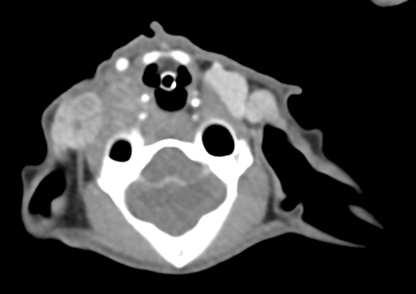
The lump turned out to be enlarged salivary glands below the right side of the lower jaw and near the ear, together with enlarged lymph nodes in the neck. However, the CT also found lesions in both lungs (small and large nodules and changes in the lung tissue) and an intussusception in the stomach, a condition in which one part of the stomach slides into another, like the sections of a telescope.

Transverse CT slice at the level of the finding

Cross-section through the upper part of the neck at the level of the abnormal salivary glands on the right.

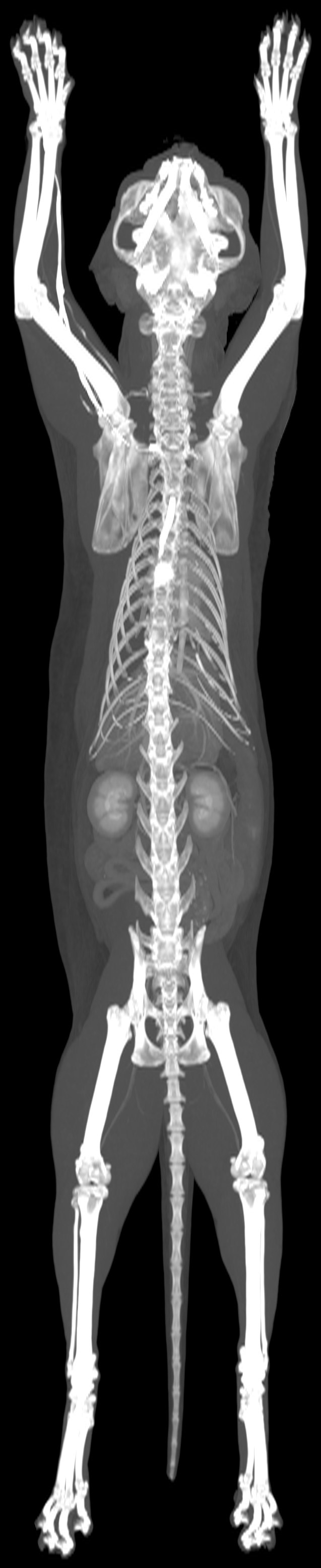
Maximum intensity projection of the whole scan

The whole scan in a single image: the skeleton, and the kidneys highlighted by the contrast agent.
Neurology, Head and neck

Seizures and walking in circles. CT found a lesion in the brain

Patient
dog, Staffordshire Bull Terrier, male, 11 years
Examination
CT of the head, pre- and post-contrast
Date
August 2026

For the past month, an eleven-year-old dog had been having epilepsy-like seizures and turning in circles to the right. The vet recommended a CT scan of the head to look for the cause in the brain and skull.

In the right hemisphere of the brain, the CT found a lesion about two centimetres in size that was pressing on its surroundings: the falx, the natural dividing wall between the hemispheres, was pushed slightly to the left. On the right, the thin bone that separates the brain from the nasal cavity was damaged, but the change had not grown into the nose. The CT also picked up breakdown of the necks of two teeth in the right upper jaw, with bone loss around one of them.

Preparation, procedure and FAQ

No food for 12 hours before the scan, water is fine. Your pet stays with us until it wakes up, and you usually go home after 3 to 5 hours. You get the radiologist's report and the images through permanent online access.

All questions and answers about CT

Staromestská 6/D,
Bratislava

Phone+421 2 2102 8700Emailvetct@vetline.skAddressStaromestská 6/D, 811 03 Bratislava
Scanning hours
Monday to Fridayby available appointments
Saturday and Sundayurgent cases, by arrangement

If you are not sure whether CT is the right examination, call us before you pay. We will tell you straight, and if a different examination would be better for the patient, we will point you to it.

Our CT unit is in the building of the Vetline veterinary hospital, which is open 24/7. With anaesthesia, this is no small detail: if something goes wrong, help is right next door, not a drive away.