Real reports translated from the Czech originals, with identifying details removed: history, technique, key findings, conclusion with differential diagnosis and recommendation. Each case also includes the version we use to explain it to the client.
Oncology, Abdomen, Head and neck
Right thyroid lobe mass and multisystemic findings in a cat treated for hyperthyroidism
Patient
cat, domestic shorthair, spayed female, 16 years
Examination
Whole-body CT, pre- and post-contrast
Date
March 2026
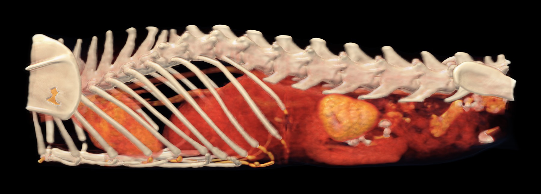
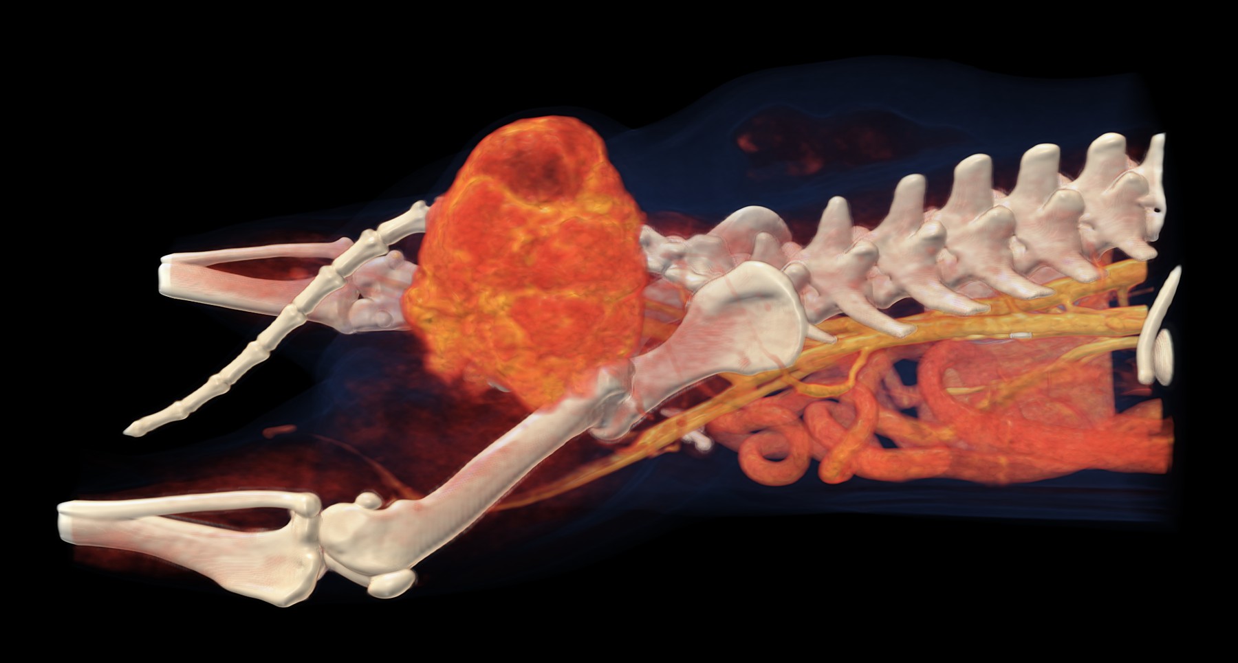
Volume rendering (VR) with an HU-based colour map from the 2 mm post-contrast series, resampled to an isotropic voxel size of 0.4 mm, left lateral view: enlarged left kidney with irregular contours, heart cranially.
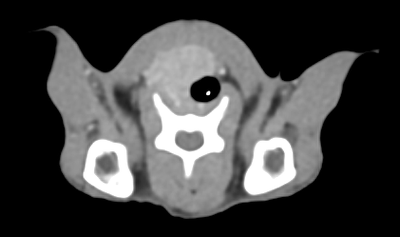
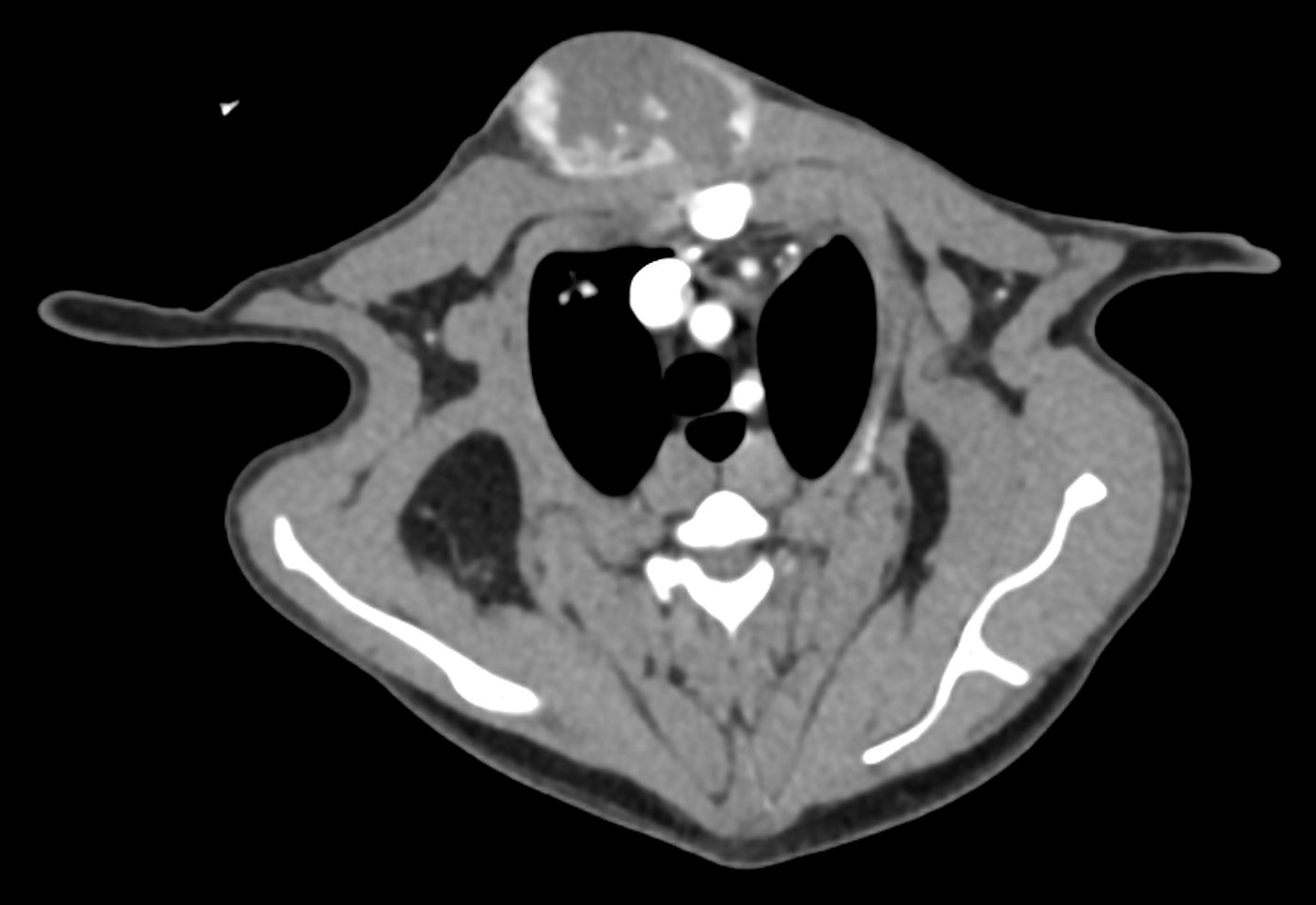
Transverse slice through the neck, post-contrast, soft tissue window W 400 / L 40: right thyroid lobe mass ventrolateral to the trachea.
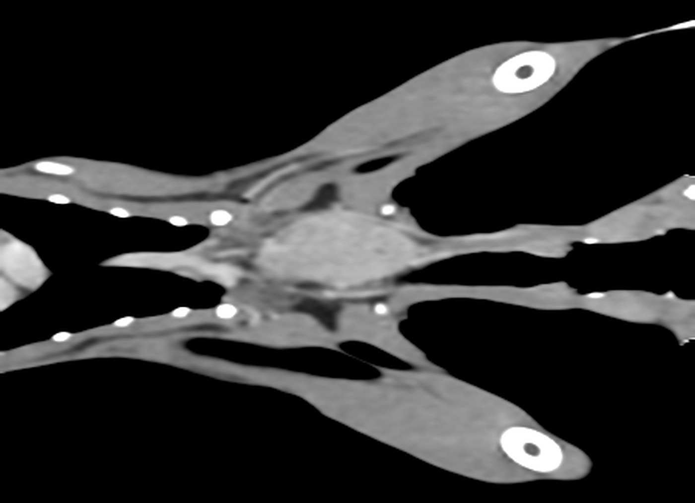
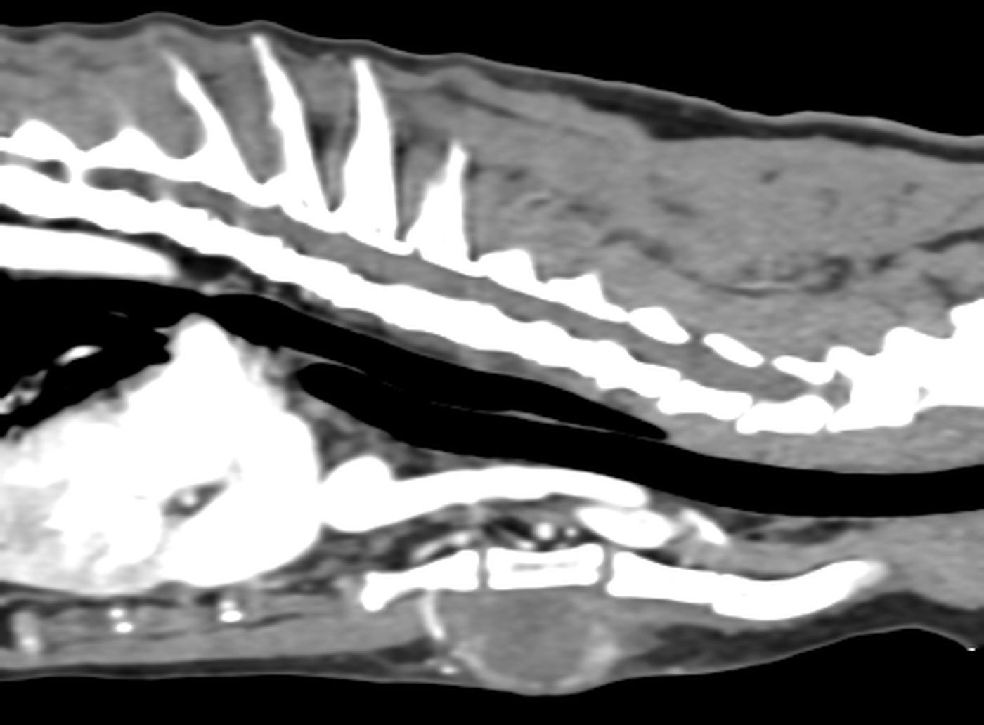
Dorsal plane through the neck at the level of the thyroid gland, post-contrast, W 400 / L 40: right thyroid lobe mass, approximately 4 cm in length.
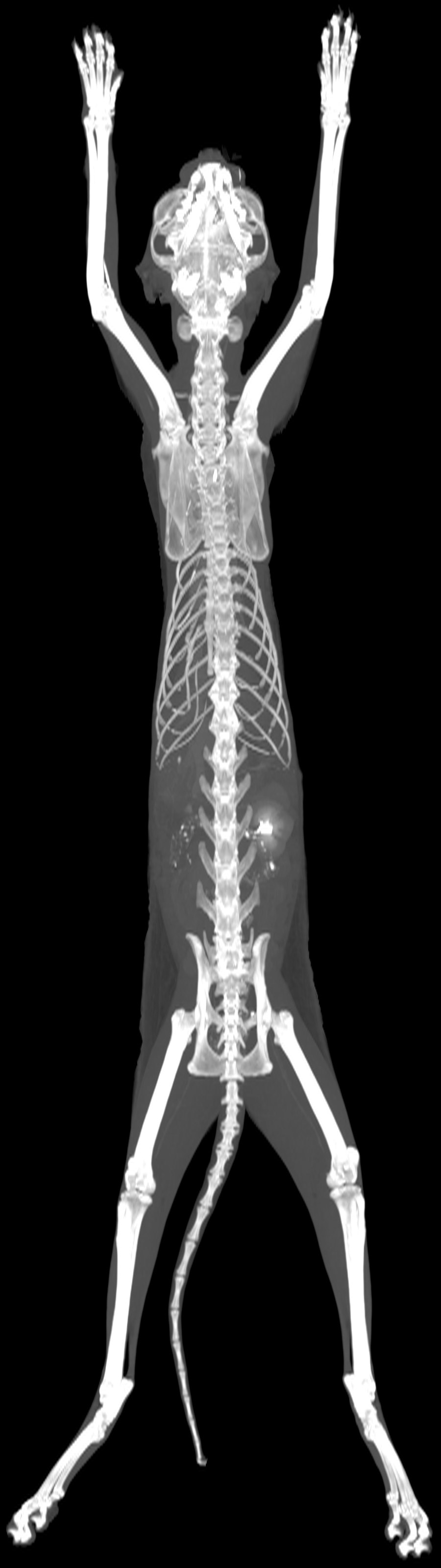
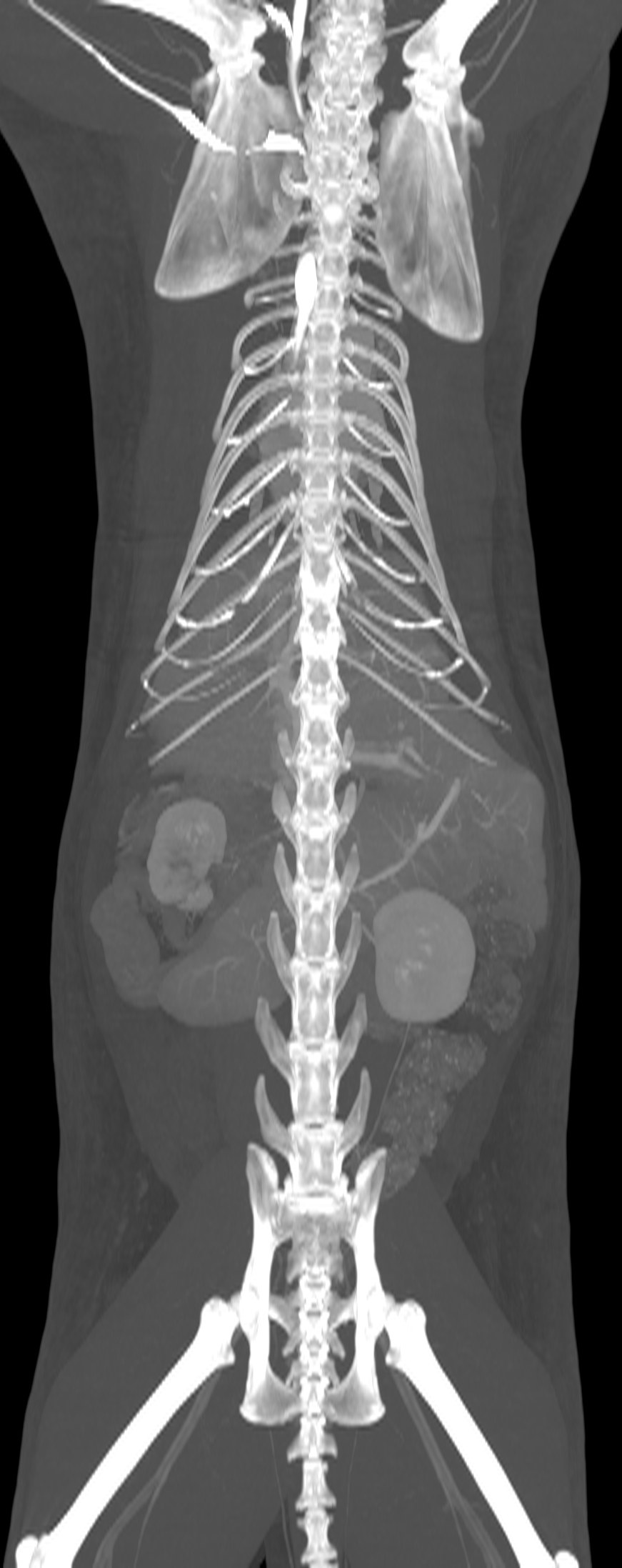
Dorsal MIP of the entire volume, post-contrast.
History
Cachexia, chronic haematemesis. Under treatment for hyperthyroidism.
Technique
4,056 slices (1 and 2 mm), pre- and post-contrast series reconstructed with medium- and high-frequency algorithms. Good study quality.
Findings
–Neck: oval, irregular mass of heterogeneous soft tissue attenuation, mildly hypoattenuating relative to the adjacent musculature, heterogeneously enhancing, located ventrally on the right, without vascular invasion or invasion of the surrounding tissues; corresponds to the right thyroid lobe (40.6 × 14.7 × 18.5 mm). Left lobe not identified.
–Thorax: enlarged sternal lymph nodes without a visible hilum. Left atrial dilatation; thickened left ventricular free wall and interventricular septum.
–Liver: hypoattenuating, non-enhancing nodular lesion in the right lateral lobe (5.5 × 7.3 × 7.8 mm); small lesion in the left medial lobe, isoattenuating on pre-contrast and hypoattenuating on post-contrast images (4.3 × 5 × 5.2 mm).
–Spleen: heterogeneous enhancement of the cranial margin; multiple irregularly shaped hypoattenuating foci.
–Kidneys: right kidney small and non-enhancing, with a triangular hyperattenuating focus at the level of the renal pelvis; left kidney enlarged, with irregular contours and heterogeneous enhancement, containing multiple oval to spherical hypoattenuating foci, some protruding beyond the renal contour.
–Stomach: diffuse circumferential wall thickening at the cardia and along the greater curvature, with homogeneous enhancement.
–Small intestine difficult to assess; multifocal fat stranding and adhesions between intestinal loops.
–Lymph nodes: enlarged gastric, splenic, cranial and caudal mesenteric, lumbar aortic, internal and medial iliac nodes, without a visible hilum.
–Peritoneum: fat stranding in the mesogastric and hypogastric regions; multifocal small volumes of free fluid.
–Ears: retention of a small amount of fluid in the right horizontal ear canal with mild thickening of its wall; multiple mineralised foci in the walls of both external ear canals (incidental dystrophic mineralisation or chronic otitis externa); reduced volume and thickened walls of both tympanic bullae, which contain material of fluid/soft tissue attenuation (common in brachycephalic dogs).
–Heart: irregular soft tissue attenuating mass at the heart base, heterogeneously enhancing, encircling the aorta, in contact with both the left and right atria and compressing the left and right pulmonary arteries (49 × 34.8 × 42.8 mm). Focal pericardial effusion on the left.
–Prostate: symmetrical, heterogeneously enhancing, with multiple small non-enhancing foci in the parenchyma.
–Pelvis: focal soft tissue thickening in the left ventral perianal region with peripheral enhancement.
–Skeleton: bilateral tarsal osteoproliferation; incomplete fusion of the ossification centres of the dorsal arch of the atlas; vertebral malformations typical of the breed.
Conclusion
–Grade 4 caudal aberrant turbinates.
–Bilateral otitis externa with accumulation of secretions in the right ear canal.
–Bilateral tympanic bulla changes consistent with a brachycephalic breed.
–Heart base mass. DDx: chemodectoma (aortic body neoplasia), haemangiosarcoma, neuroendocrine neoplasia.
Right-sided mandibular and parotid sialoadenopathy, multifocal pneumopathy and gastrogastric intussusception in a cat
Patient
cat, British Shorthair, neutered male, 11 years
Examination
Whole-body CT, pre- and post-contrast
Date
March 2026
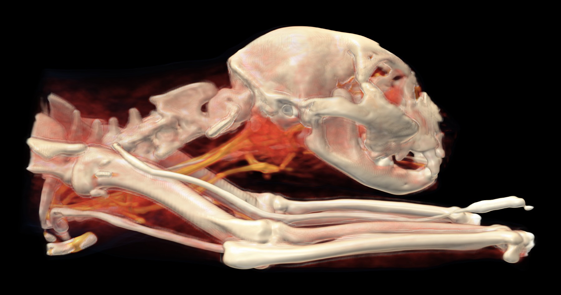
Volume rendering (VR) with an HU-based colour map from the 2 mm post-contrast series, resampled to an isotropic voxel size of 0.4 mm, right lateral view: region of right-sided sialoadenopathy and cervical lymphadenopathy, cervical vessels; along the thoracic limb, the cannula extension line containing contrast medium.
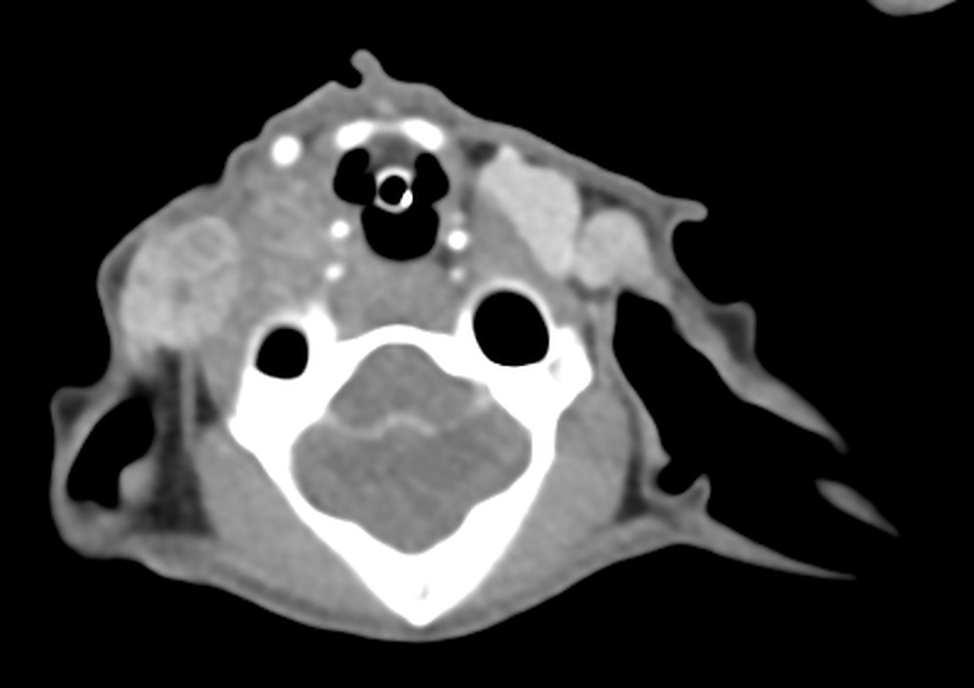
Transverse slice at the level of the skull base, post-contrast, W 400 / L 40: region of right-sided mandibular and parotid sialoadenopathy.
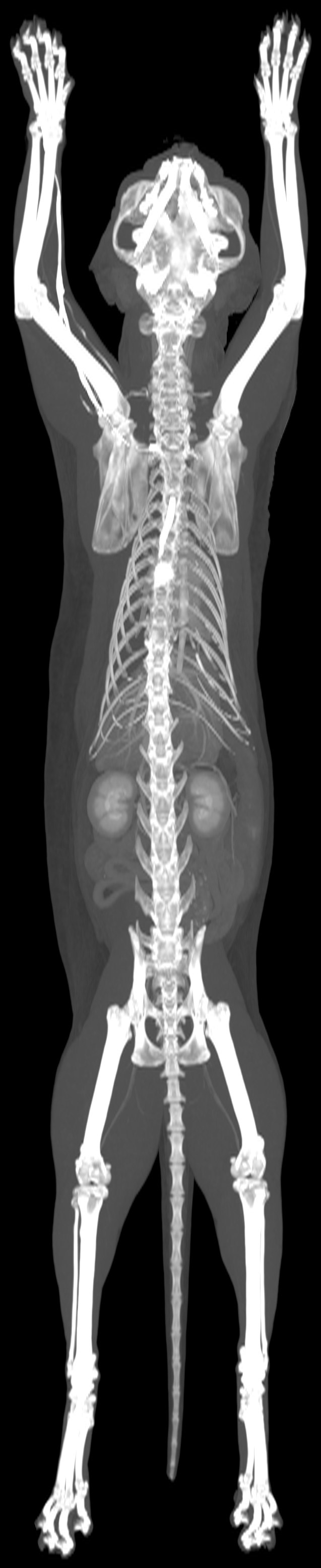
Dorsal MIP of the entire volume, post-contrast.
History
Firm mass on the neck for approximately 2 months. Pruritus of the neck and head, self-trauma.
Technique
3,811 slices (1 and 2 mm), pre- and post-contrast series reconstructed with medium- and high-frequency algorithms. Good study quality.
Findings
–Salivary glands: right mandibular and parotid glands diffusely enlarged, irregular in shape, of soft tissue attenuation, heterogeneously enhancing and poorly demarcated from the surrounding tissues.
–Lymph nodes of the head and neck: enlarged right medial retropharyngeal (spherical, heterogeneously enhancing), right parotid, bilateral ventral superficial cervical and left dorsal superficial cervical nodes, without a visible hilum.
–Lungs: small and large irregularly shaped nodules of soft tissue attenuation in all lobes, the large ones heterogeneously enhancing; multifocal ground-glass opacities, both central and subpleural; parenchymal bands.
–Heart: thickened left ventricular free wall and interventricular septum; left atrial dilatation.
–Stomach: small and spherical; cardia and fundus displaced aborally into the lumen of the gastric body, with concentric wall layering and interposed fat (intussusception); diffuse wall thickening of the gastric body (intussuscipiens); pylorus in a normal position; no mural mass or nodule.
Conclusion
–Right mandibular and parotid sialoadenopathy. DDx: sialoadenitis, neoplasia.
Biopsy and correlation with cytology or histopathology.
Note
Gastrogastric intussusception may be transient, with spontaneous resolution. The thickening of the outer wall layer (intussuscipiens) is probably a consequence of it, but may also reflect an inflammatory or neoplastic process. Gastric ultrasound is recommended.
Translated from the Czech original, with identifying details removed. Report signed by MVDr. Ladislav Stehlík, Ph.D..
Bilateral chylous pleural effusion with atelectasis; psoas minor thickening in a cat following excision of an adenocarcinoma
Patient
cat, domestic shorthair, spayed female, 9 years
Examination
Whole-body CT, pre- and post-contrast
Date
May 2026
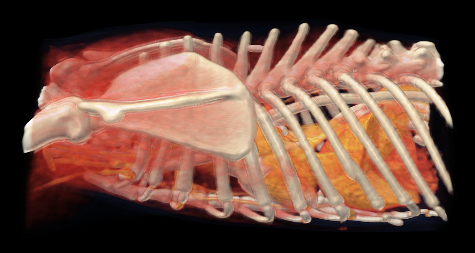
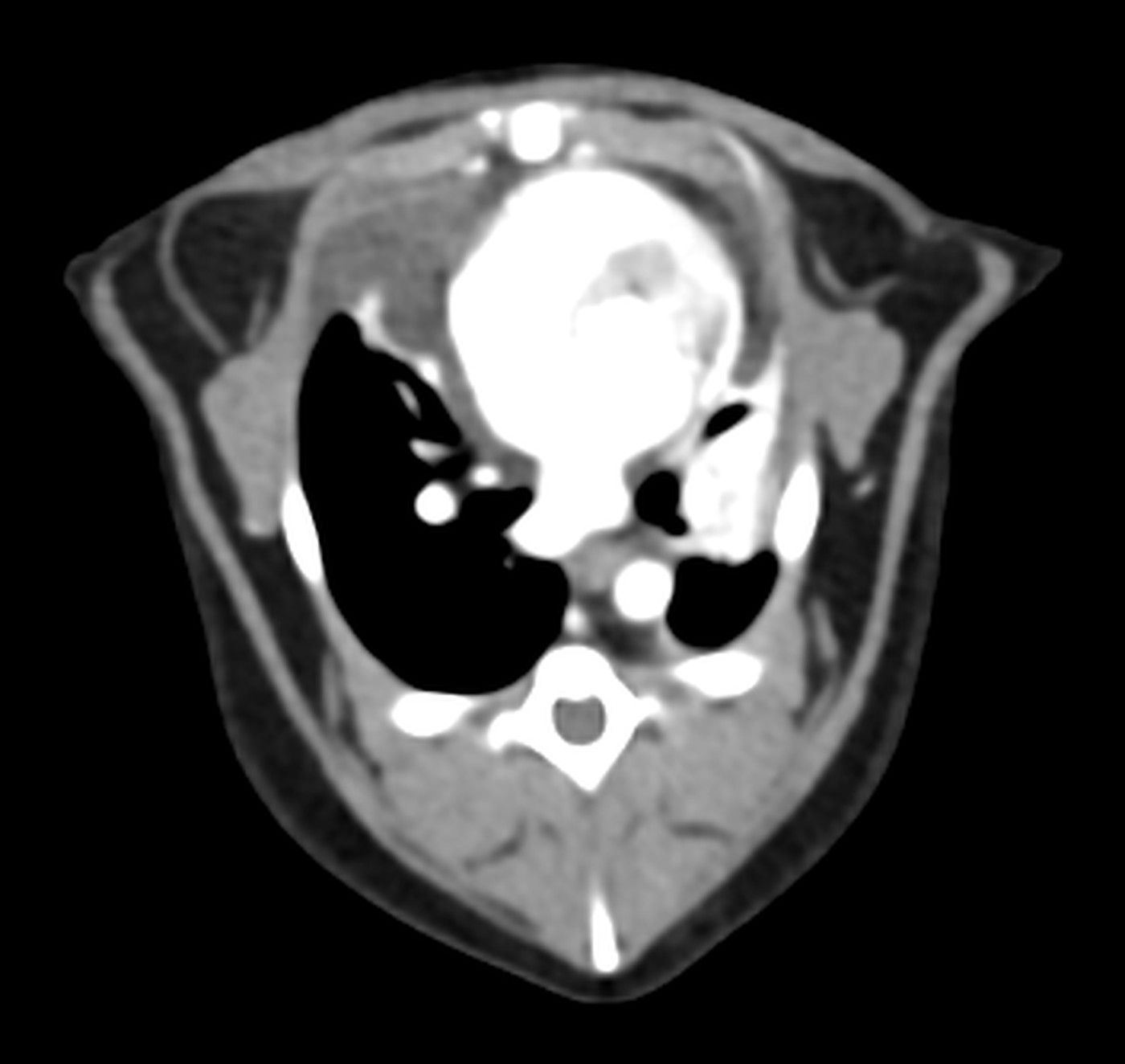
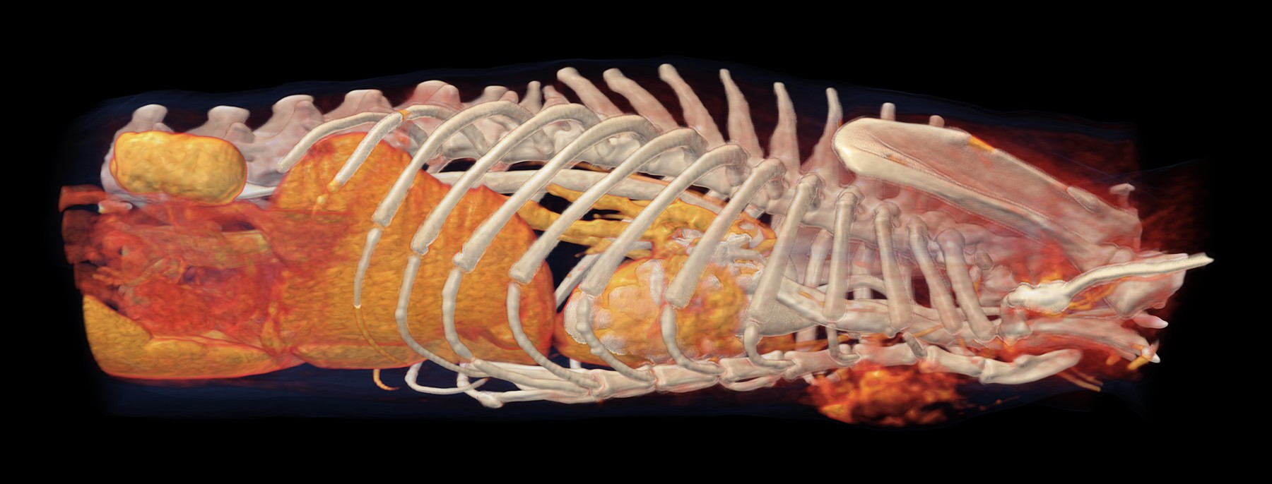
Volume rendering (VR) with an HU-based colour map from the 2 mm post-contrast series, resampled to an isotropic voxel size of 0.4 mm, left lateral view of the thorax. The pleural effusion is of fluid attenuation and is not displayed in VR; it is assessed on the transverse slices.
Transverse slice through the thorax, post-contrast, W 400 / L 40: bilateral pleural effusion, lobar atelectasis.
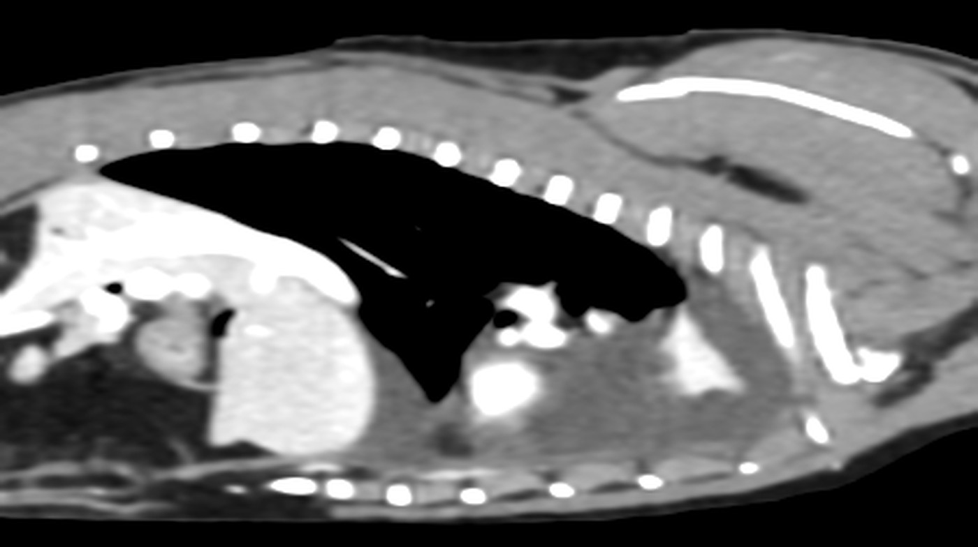
Sagittal plane through the thorax, post-contrast, W 400 / L 40: pleural effusion and collapsed lung lobes.
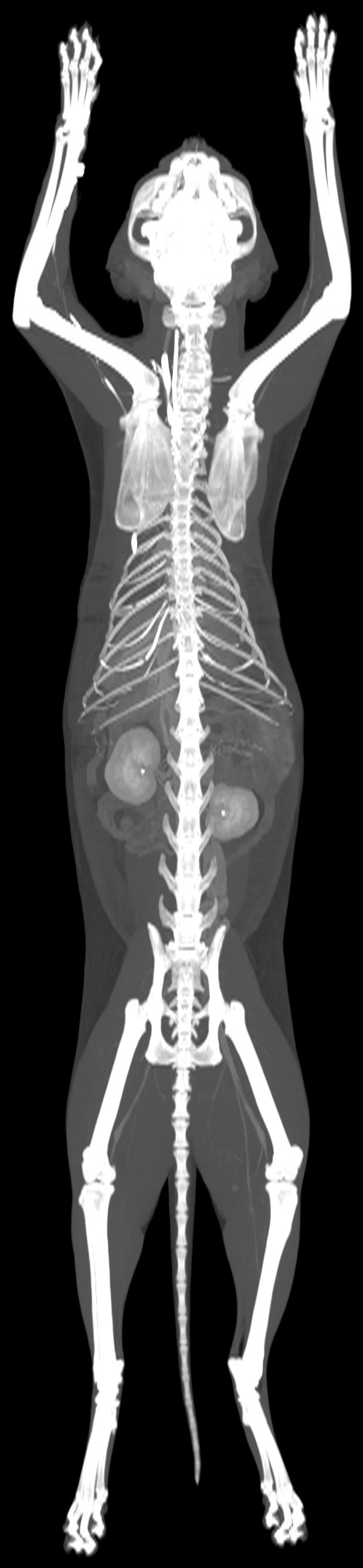
Dorsal MIP of the entire volume, post-contrast.
History
Excision of a thigh mass 2 months previously (adenocarcinoma). Laparotomy for suspected diaphragmatic hernia 5 days before CT. Currently tachypnoea, abdominal breathing, muffled heart sounds and elevated renal parameters. Approximately 100 ml of chylous fluid drained from the thoracic cavity before CT.
Technique
4,433 slices (1 and 2 mm), pre- and post-contrast series reconstructed with medium- and high-frequency algorithms. Good study quality.
Findings
–Lungs: right cranial and right middle lobes, both the cranial and caudal parts of the left cranial lobe, and the accessory lobe consolidated with reduced volume and air-filled lobar bronchi; caudal lobes aerated.
–Pleura: fluid in both pleural spaces; multifocal thickening of the visceral pleura, without nodules or masses.
–Lymph nodes: enlarged cranial mediastinal nodes (irregular, spherical, heterogeneously enhancing) and mildly enlarged sternal nodes; enlarged left popliteal node.
–Adrenal glands: symmetrically enlarged (DV right 5.2 mm, left 5.1 mm), with normal shape and enhancement and no vascular invasion.
–Kidneys: mild multifocal surface indentations at the poles; small bilateral mineralisations in the renal pelves.
–Muscles: bilateral asymmetrical thickening of the hypaxial musculature at the level of L7 and the sacrum, in the location of the psoas minor muscles, heterogeneously enhancing, without vascular invasion; focal heterogeneous enhancement of the soft tissues dorsomedial to the body of the right ilium.
–Peritoneum: multiple small gas bubbles and diffuse mild fat stranding.
Conclusion
–Bilateral pleural effusion.
–Atelectasis of several lung lobes, most likely secondary to the effusion.
Ventral thoracic wall mass with probable invasion of the deep pectoral muscle; suspected ectopic ureters
Patient
cat, Maine Coon, male, 10 years
Examination
CT of the thorax, abdomen and pelvis, pre- and post-contrast
Date
June 2026
Volume rendering (VR) with an HU-based colour map from the 2 mm post-contrast series (late venous phase), resampled to an isotropic voxel size of 0.4 mm, right lateral view: ventral thoracic wall mass at the level of the cranial sternebrae; kidneys and liver post-contrast.
Transverse slice at the level of the cranial sternebrae, post-contrast, W 400 / L 40: heterogeneously enhancing ventral thoracic wall mass.
Midsagittal plane, post-contrast, W 400 / L 40: ventral thoracic wall mass at the level of the cranial sternebrae.
Dorsal MIP of the entire volume, post-contrast.
History
Mass on the ventral thoracic wall adjacent to the sternum.
Technique
2,457 slices (1 and 2 mm), pre- and post-contrast series reconstructed with medium- and high-frequency algorithms; 2 post-contrast series in the late venous and excretory phases. Good study quality; motion artefacts in the lungs.
Findings
–Thoracic wall: spherical, heterogeneously enhancing mass of soft tissue attenuation in the subcutis of the ventral thoracic wall at the level of sternebrae 1–3 (49.5 × 22.8 × 35.6 mm); focal invasion of the left deep pectoral muscle in the median plane cannot be excluded; very close contact with the right deep pectoral muscle; no invasion of the thoracic cavity or pleural spaces.
–Lungs: right middle lobe and caudal part of the left cranial lobe with reduced volume and consolidation, air-filled lobar bronchi; remaining lobes difficult to assess owing to motion artefacts.
–Heart: dilatation of the left atrium and left ventricle.
–Kidneys and ureters: right kidney small, with irregular contours and heterogeneous enhancement; left kidney unremarkable; focal dilatation of the left ureter in its proximal third; exact site of insertion of both ureters not identified; urinary bladder distended, containing a very small amount of contrast medium; urethra filled with contrast medium.
Conclusion
–Ventral thoracic wall mass, very likely with focal invasion of the left deep pectoral muscle. Most likely neoplasia (suspected sarcoma).
–Right-sided nephropathy.
–Focal left hydroureter.
–Very likely ectopic insertion of both ureters into the proximal urethra.
Recommendation
Biopsy and correlation with cytology or histopathology. Ultrasound of the urinary tract. To confirm or rule out ectopic ureters, repeat CT (excretory CT urography) after an enema and with an appropriately filled urinary bladder (avoiding both overdistension and complete emptying).
Translated from the Czech original, with identifying details removed. Report signed by MVDr. Ladislav Stehlík, Ph.D..
Oncological staging of a soft tissue sarcoma of the pelvic region
Patient
dog, mixed breed, spayed female, 11 years
Examination
Whole-body CT, pre- and post-contrast
Date
August 2026
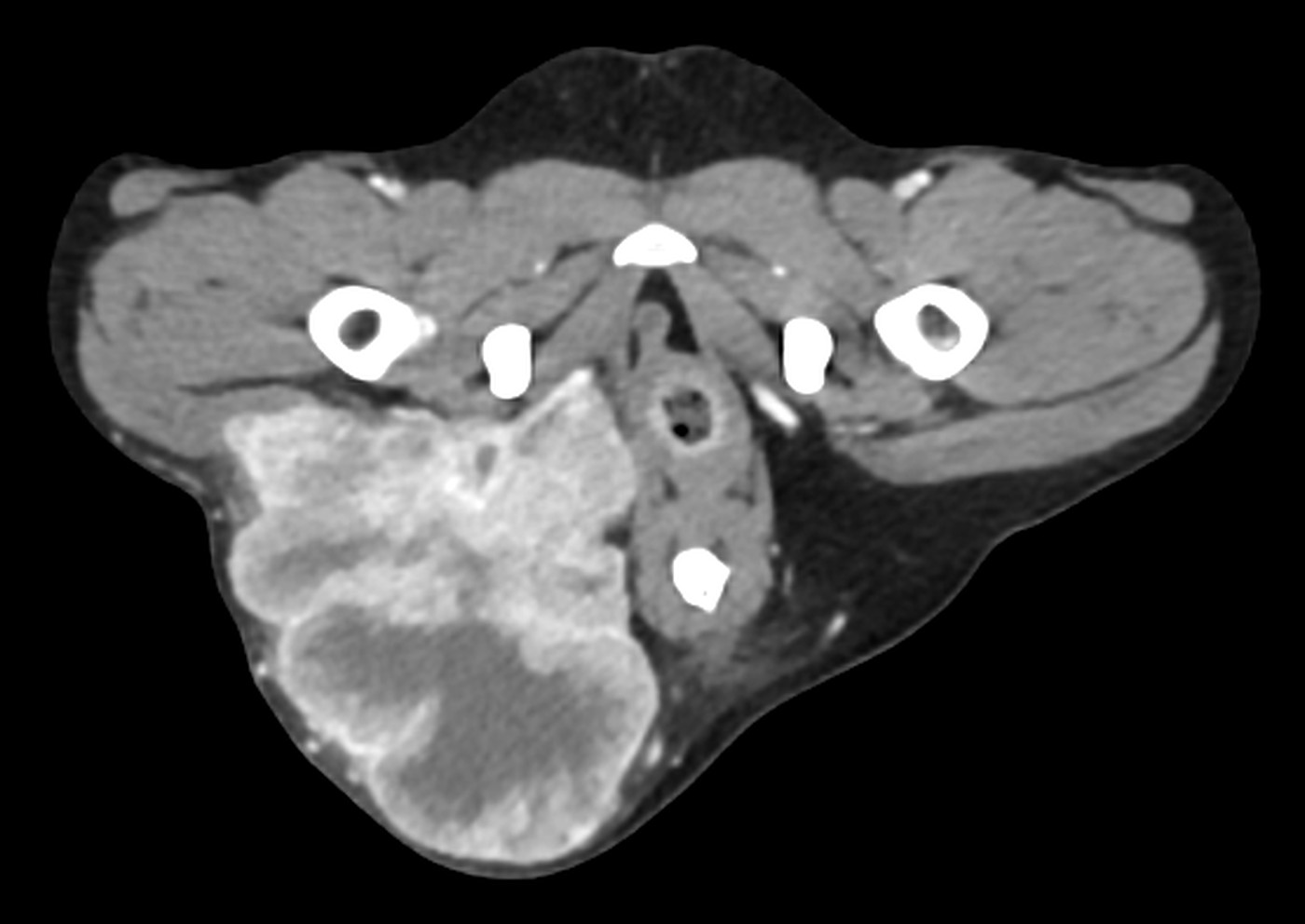
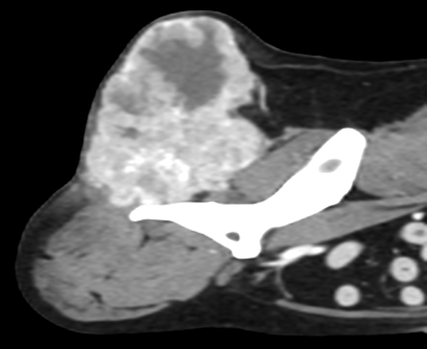
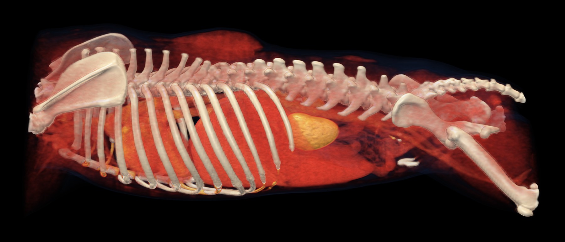
Volume rendering (VR) with an HU-based colour map from the 2 mm post-contrast series, resampled to an isotropic voxel size of 0.4 mm, pelvic and lumbar region, right lateral view: multilobular mass dorsal to the right hip joint, aorta and iliac arteries; a second mass shows through in the contralateral lumbar region.
Transverse slice at the level of the hip joints, post-contrast, W 400 / L 40: multilobular, heterogeneously enhancing mass dorsally on the right, with displacement of the rectum to the left.
Parasagittal plane through the mass on the right, post-contrast, W 400 / L 40: multilobular, heterogeneously enhancing mass dorsal to the right hip joint.
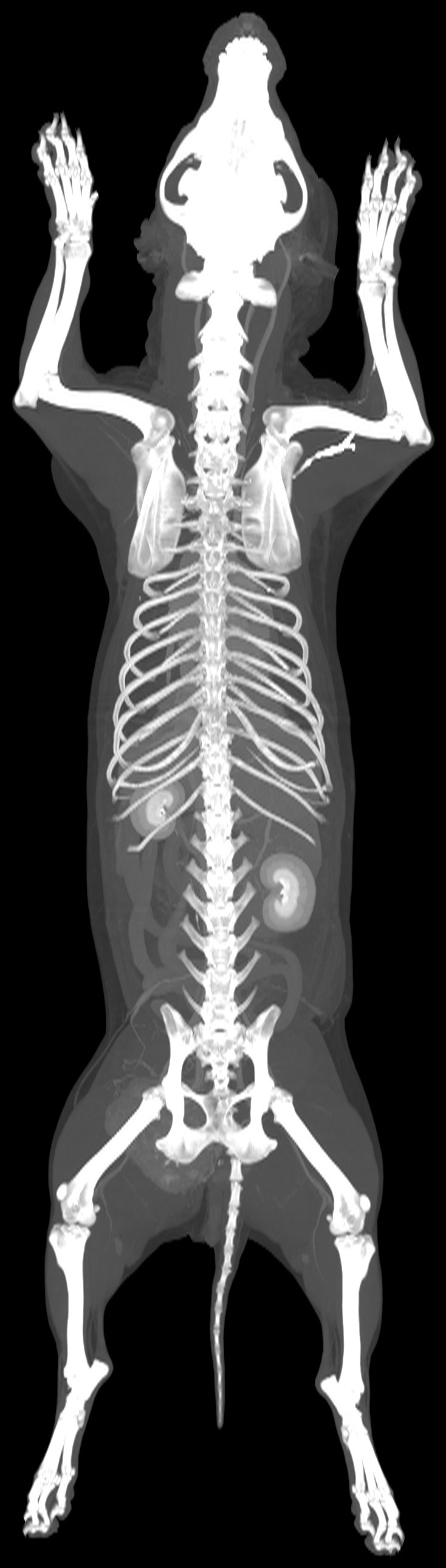
Dorsal MIP of the entire volume, post-contrast.
History
Mass at the level of the pelvis on the right; biopsy result: soft tissue sarcoma. Oncological screening.
Technique
3,438 slices (1 and 2 mm), pre- and post-contrast series reconstructed with medium- and high-frequency algorithms; 1 post-contrast series in the late venous phase. Excellent study quality.
Findings
–Pelvis: multilobular mass of soft tissue attenuation with central mineralisation, heterogeneously enhancing and well defined, located dorsally at the level of the right hip joint (69 × 69 × 64.5 mm); invasion of the right gluteal musculature, with suspected invasion also of the right piriformis, biceps femoris and semitendinosus muscles; extends into the pelvic cavity from the right dorsal aspect; mild compression and displacement of the rectum to the left; adjacent bone unremarkable.
–Left lumbar region: irregular, elongated mass of soft tissue attenuation with heterogeneous enhancement, located subfascially within the fat from the level of L5 to the wing of the ilium (68 × 20 × 51 mm), well defined.
–Lungs: multiple miliary mineralised nodules, predominantly subpleural; small thin-walled, air-filled lesion in the right caudal lobe (d = 6 mm).
–Anal sacs: subtle mineralised sediment (sacculoliths); mild compression of the right anal sac due to the mass effect.
Conclusion
–Multilobular expansile and invasive mass at the level of the right side of the pelvis, consistent with neoplasia, most likely sarcoma.
dog, Jack Russell Terrier, spayed female, 10 years
Examination
Whole-body CT, pre- and post-contrast
Date
May 2026
Volume rendering (VR) with an HU-based colour map from the 2 mm post-contrast series, resampled to an isotropic voxel size of 0.4 mm, left lateral view. The lymphadenopathy and subcutaneous masses are assessed on the cross-sectional images.
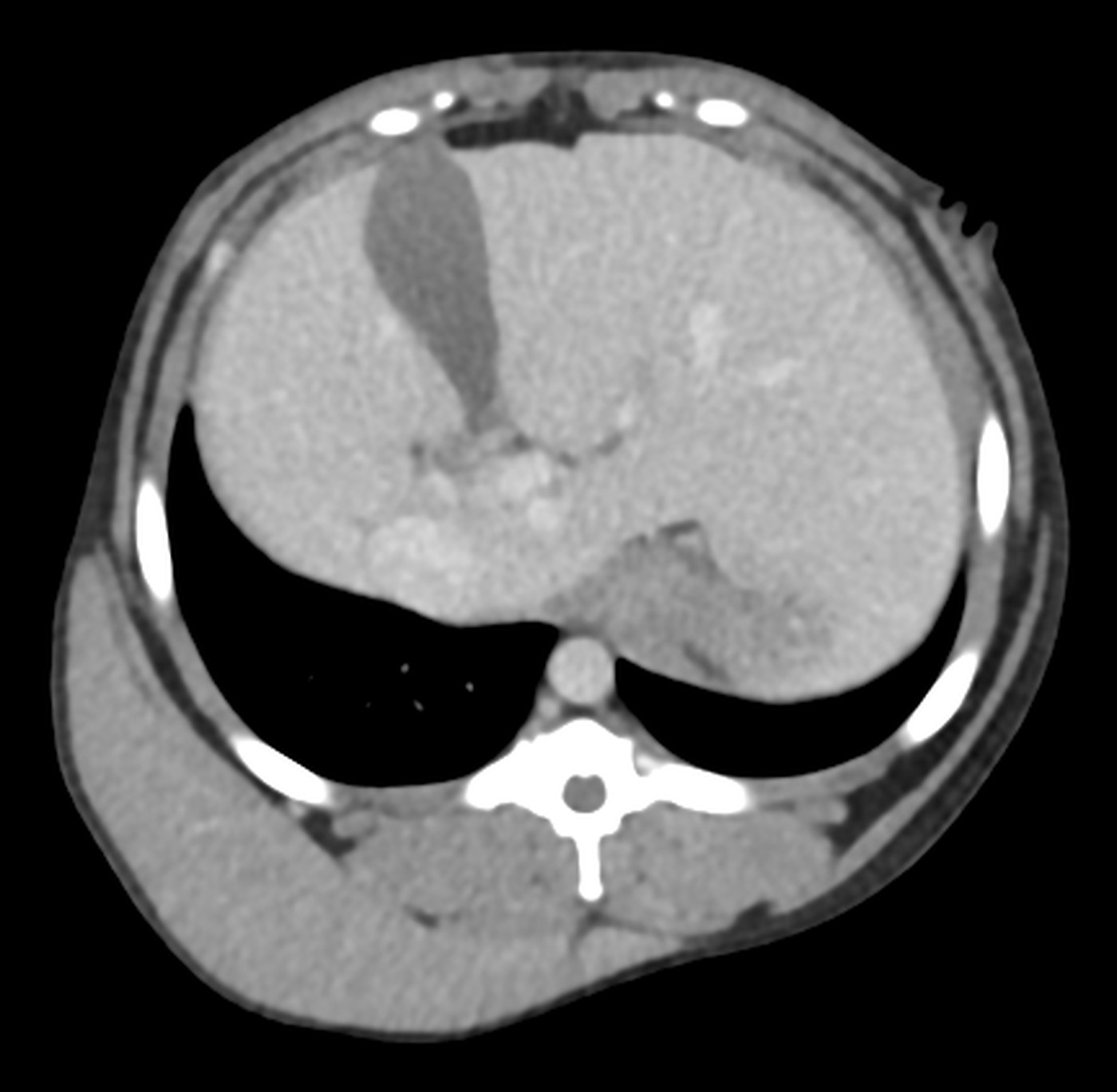
Transverse slice through the cranial abdomen, post-contrast, W 400 / L 40: hepatomegaly, plaque-like subcutaneous masses dorsally.
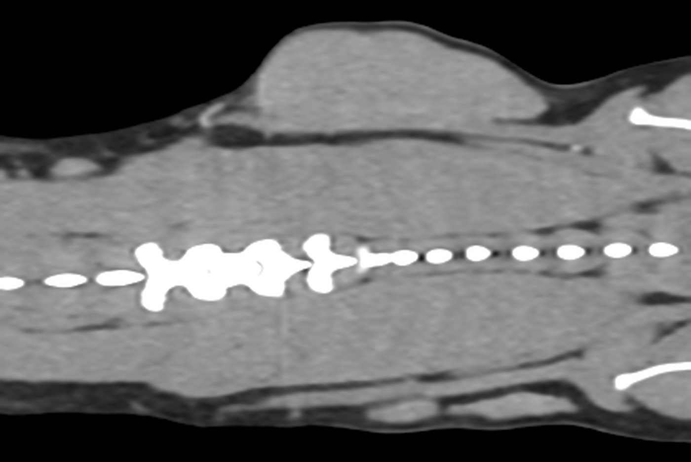
Dorsal plane through the trunk, post-contrast, W 400 / L 40: subcutaneous thoracic wall mass.
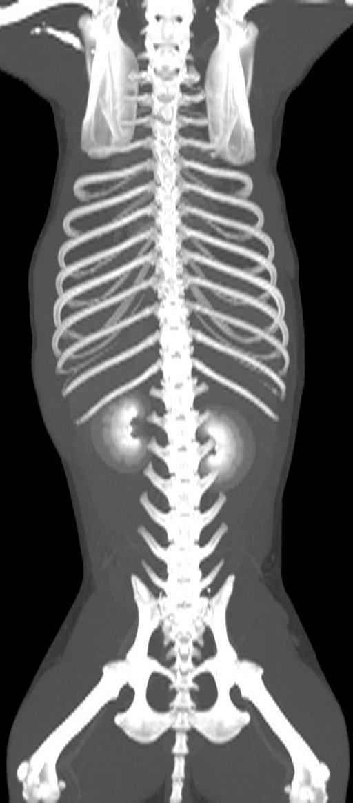
Dorsal MIP of the entire volume, post-contrast.
History
Gastrointestinal signs for approximately one year. Currently generalised lymphadenopathy, hyperthermia and a large subcutaneous mass on the right side of the thorax.
Technique
3,159 slices (1 and 2 mm), pre- and post-contrast series reconstructed with medium- and high-frequency algorithms; 2 post-contrast series in the late venous phase. Excellent study quality.
Findings
–Lymph nodes: enlarged, without a visible hilum, homogeneous; mandibular, parotid and medial retropharyngeal bilaterally, superficial and deep cervical, sternal, cranial mediastinal, axillary, iliosacral, superficial inguinal and popliteal bilaterally.
–Subcutis and superficial fascia of the thoracic and abdominal walls: multifocal thickening ranging to plaque-like masses of soft tissue attenuation with homogeneous enhancement, bilaterally and dorsally, well defined, without intermuscular spread; the largest is dorsolateral on the right, extending from T6 to L3–4.
–Thymus: enlarged, homogeneously enhancing.
–Liver and spleen: diffusely enlarged, homogeneously enhancing, with smooth margins.
–Brain: asymmetry of the lateral ventricles (right > left).
Conclusion
–Generalised lymphadenopathy.
–Bilateral subcutaneous masses of the thoracic and abdominal walls.
–Most likely DDx: lymphoma, disseminated histiocytic sarcoma, reactive histiocytosis; among infectious diseases, Leishmania sp. (travel to or residence in an endemic area must be confirmed).
Recommendation
Biopsy and correlation with cytology or histopathology, with additional specialised tests where appropriate (Leishmania PCR, PARR, immunohistochemistry).
Translated from the Czech original, with identifying details removed. Report signed by MVDr. Ladislav Stehlík, Ph.D..
4,132 slices (1 and 2 mm), pre- and post-contrast series reconstructed with medium- and high-frequency algorithms; 2 post-contrast series of the abdomen in the late and excretory phases. Good study quality; motion artefacts in the lungs.
Findings
–Liver: diffusely mildly enlarged, with rounded margins, normal attenuation and homogeneous enhancement; surrounding fat unremarkable. Gallbladder and common bile duct unremarkable.
–Lymph nodes: hepatic nodes enlarged, spherical, without a visible hilum, of heterogeneous soft tissue and fat attenuation, heterogeneously enhancing.
–No abnormalities detected in the lungs; several missing teeth.
Biopsy and correlation with cytology or histopathology.
Note
Diffuse hepatomegaly is a non-specific finding that may accompany degenerative hepatopathies and hepatitis of various types, but also neoplasia, most commonly round cell or other infiltrative neoplasms.
Translated from the Czech original, with identifying details removed. Report signed by MVDr. Ladislav Stehlík, Ph.D..
6,205 slices (1 and 2 mm), pre- and post-contrast series reconstructed with medium- and high-frequency algorithms; 2 post-contrast series in the late venous and excretory phases. Good study quality; motion artefacts.
Findings
–Abdominal cavity: no detectable abnormality of the pancreas or other organs; adrenal glands within normal limits (DV right 5–6 mm, left 7–7.3 mm).
–Lungs: multifocal ground-glass opacities with an irregular distribution in several lobes.
–Heart: dilatation of the left atrium and left ventricle.
–Spine: focal osteoproliferation of the L7 vertebral body on the left; mild narrowing of the left L7–S1 neuroforamen.
Conclusion
–No detectable abnormality of the pancreas or other abdominal organs.
–The pulmonary changes may be secondary to anaesthesia and inadequate lung inflation.
–Mild left L7–S1 foraminal stenosis.
Recommendation
Triple-phase CT angiography of the abdomen.
Translated from the Czech original, with identifying details removed. Report signed by MVDr. Ladislav Stehlík, Ph.D..
Left elbow dysplasia: medial coronoid disease (MCD) without fragmentation
Patient
male, 5 years
Examination
CT of the forelimbs, pre- and post-contrast
Date
September 2026
History
Recurrent left thoracic limb lameness for approximately 1 year.
Technique
1,268 slices (1 and 3 mm), pre- and post-contrast series reconstructed with medium- and high-frequency algorithms; scans and reconstructions with a large FOV. Good study quality.
Findings
–Left elbow: well-defined but poorly marginated medial coronoid process of heterogeneous attenuation; normal humeroradioulnar congruity; anconeal process unremarkable; no evidence of new bone formation; articular and periarticular soft tissues without abnormalities.
–Long bones, other joints and right thoracic limb: no abnormalities detected.
Conclusion
–Left elbow dysplasia characterised by medial coronoid disease (MCD) without fragmentation.
Translated from the Czech original, with identifying details removed. Report signed by MVDr. Ladislav Stehlík, Ph.D..
Nodular lesion of the right cerebral hemisphere with destruction of the cribriform plate in a dog with epileptiform seizures
Patient
dog, Staffordshire Bull Terrier, male, 11 years
Examination
CT of the head, pre- and post-contrast
Date
August 2026
History
Epileptiform seizures and circling to the right over the past month.
Technique
1,202 slices (1 and 2 mm), pre- and post-contrast series reconstructed with medium- and high-frequency algorithms. Excellent study quality.
Findings
–CNS: heterogeneous pre-contrast attenuation of the rostral part of the right hemisphere and the olfactory lobe; heterogeneous, predominantly peripheral enhancement in the central part of the right hemisphere (23 × 17 × 12 mm); focal displacement of the falx cerebri to the left; asymmetry of the lateral ventricles (left > right), with contents of fluid attenuation; focal destruction of the right cribriform plate without extension into the nasal cavity.
–Teeth: multifocal destruction/resorption at the level of the necks of teeth 105 and 106; focal osteolysis of the alveolar bone of tooth 106; no communication with the nasal cavity.
Conclusion
–Nodular lesion in the right cerebral hemisphere. DDx: neoplasia (suspected glioma), abscess.
–Destruction of the right cribriform plate, suspected to be secondary to the brain lesion (inflammatory or neoplastic activity), or possibly pressure necrosis of the bone (oedema, raised intracranial pressure).
Recommendation
Neurological examination, MRI of the brain and CSF analysis.
Translated from the Czech original, with identifying details removed. Report signed by MVDr. Ladislav Stehlík, Ph.D..
3,223 slices (1 and 2 mm), pre- and post-contrast series reconstructed with medium- and high-frequency algorithms; 3 post-contrast series in the late venous phase. Diagnostic study quality; motion artefacts in the lungs.
Findings
–Lungs: multiple small nodules of soft tissue attenuation, both central and subpleural, spherical and irregular, some poorly defined (halo sign).
–Mediastinum: small structure of metallic attenuation with marked artefacts, dorsal to the oesophagus and ventral to the aorta; oesophageal lumen unremarkable.
–Lymph nodes: enlarged cranial mediastinal, sternal, hepatic, gastric, splenic, jejunal and lumbar aortic nodes, without a visible hilum.
–Heart: dilatation of both atria and both ventricles.
–Liver: diffusely enlarged, with rounded margins, multifocal heterogeneous enhancement and a periportal halo. Gallbladder distended, with a hyperattenuating peripheral rim of irregular width and a small amount of mineralised sediment.
–Spleen not visualised (previous splenectomy).
–Kidneys: both enlarged, irregular in shape and heterogeneously enhancing, with multiple parenchymal nodules to masses of soft tissue attenuation, homogeneously enhancing, hypoattenuating relative to the cortex and protruding beyond the renal contour.
–Ureters: left ureter unremarkable, with normal filling; right ureter multifocally thickened and heterogeneously enhancing, with no contrast filling in any of the post-contrast series.
–Peritoneum: multifocal small volumes of free fluid and fat stranding.
Conclusion
–Multifocal lymphadenopathy, most likely neoplastic. DDx: reactive.
–Hepatopathy, nephropathy, right ureteral thickening; the most likely common cause is lymphoma.
–Pulmonary nodules consistent with metastases.
–Cholecystopathy. DDx: chronic cholecystitis, lymphoma, other neoplasia.
–Peritonitis.
–Generalised cardiomegaly.
Recommendation
Biopsy and correlation with cytology or histopathology.
Translated from the Czech original, with identifying details removed. Report signed by MVDr. Ladislav Stehlík, Ph.D..
Hepatosplenomegaly, multifocal lymphadenopathy and pneumonia in an anaemic dog
Patient
dog, German Shepherd Dog, spayed female, 11 years
Examination
CT of the neck, thorax, abdomen and pelvis, pre- and post-contrast
Date
August 2026
History
Dyspnoea, lethargy, anaemia.
Technique
4,010 slices (1 and 2 mm), pre- and post-contrast series reconstructed with medium- and high-frequency algorithms; 3 post-contrast series in the late venous and excretory phases. Diagnostic study quality; motion artefacts.
Findings
–Neck: enlarged left superficial cervical lymph node, without a visible hilum, with homogeneous enhancement.
–Lungs: multiple miliary and small nodules of soft tissue attenuation, diffusely distributed; consolidation of the ventral third of the right cranial lobe and of the cranial part of the left cranial lobe with air bronchograms; in the remaining lobes, ground-glass opacities ranging to consolidation with a diffuse distribution.
–Thoracic lymph nodes: enlarged left axillary, sternal, one cranial mediastinal and the tracheobronchial nodes, without a visible hilum, some with heterogeneous enhancement.
–Liver and gallbladder: diffuse hepatomegaly with rounded, smooth margins; thickened gallbladder wall with a peripheral rim of fluid attenuation (‘sandwich’ appearance); mineralised sediment.
–Spleen: diffuse splenomegaly with homogeneous enhancement.
–Abdominal lymph nodes: enlarged hepatic (spherical), splenic, lumbar aortic and medial iliac nodes, with heterogeneous enhancement.
–Peritoneum: multifocal free fluid.
–Skeleton: bilateral osteoproliferation of the femoral heads and acetabula; multiple osteoproliferations of the lumbar vertebral bodies, with narrowing of the right neuroforamen at L7–S1.
Conclusion
–Diffuse hepatomegaly and splenomegaly, multifocal lymphadenopathy. DDx: lymphoma, systemic infection, extramedullary haematopoiesis (the splenomegaly alone may also be secondary to the anaemia).
–Gallbladder wall thickening consistent with oedema. DDx: anaphylaxis, cholecystitis, hypoalbuminaemia.
–Small-volume ascites. DDx: neoplastic, secondary to hepatopathy.
–Pulmonary consolidation with this distribution is most consistent with pneumonia.
–Pulmonary ground-glass opacities. DDx: lymphoma, interstitial pneumopathy, pneumonia; they may also result from motion artefacts or anaesthesia.
Incidental findings
–Spondylosis deformans.
–Degenerative lumbosacral stenosis (DLSS).
Recommendation
Biopsy and correlation with cytology or histopathology.
Translated from the Czech original, with identifying details removed. Report signed by MVDr. Ladislav Stehlík, Ph.D..