Real reports translated from the Czech originals, with identifying details removed: history, technique, key findings, conclusion with differential diagnosis and recommendation. Each case also includes the version we use to explain it to the client.
Oncology, Abdomen, Head and neck
Right thyroid lobe mass and multisystemic findings in a cat treated for hyperthyroidism
Patient
cat, domestic shorthair, spayed female, 16 years
Examination
Whole-body CT, pre- and post-contrast
Date
March 2026
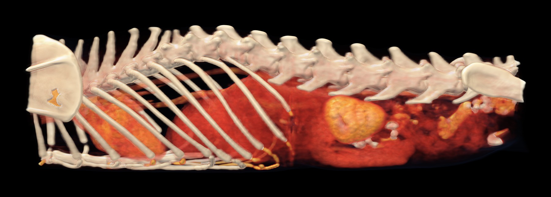
Volume rendering (VR) with an HU-based colour map from the 2 mm post-contrast series, resampled to an isotropic voxel size of 0.4 mm, left lateral view: enlarged left kidney with irregular contours, heart cranially.
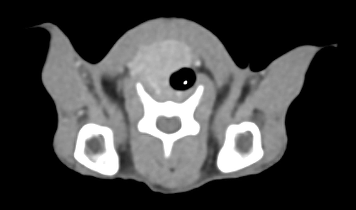
Transverse slice through the neck, post-contrast, soft tissue window W 400 / L 40: right thyroid lobe mass ventrolateral to the trachea.
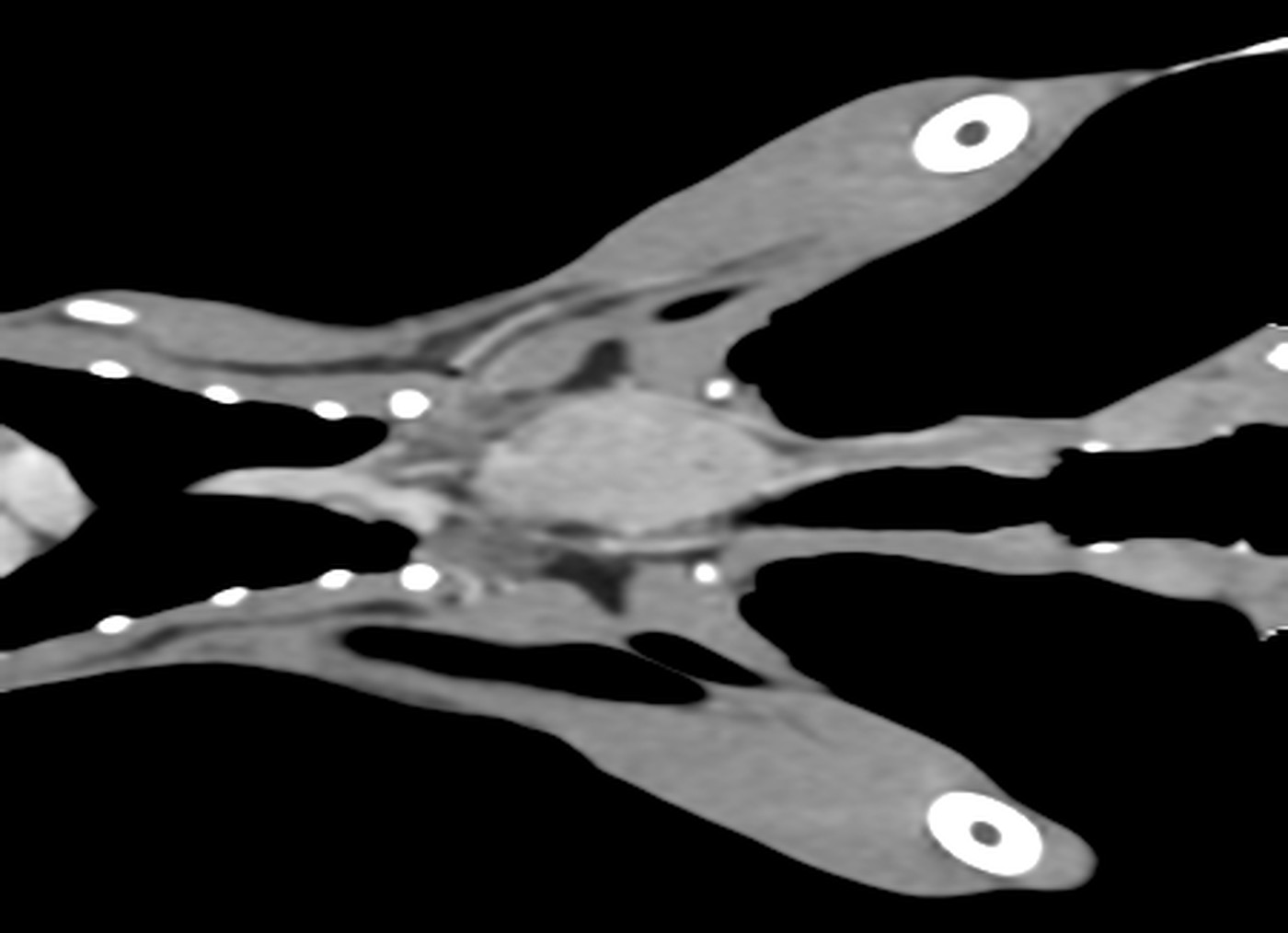
Dorsal plane through the neck at the level of the thyroid gland, post-contrast, W 400 / L 40: right thyroid lobe mass, approximately 4 cm in length.
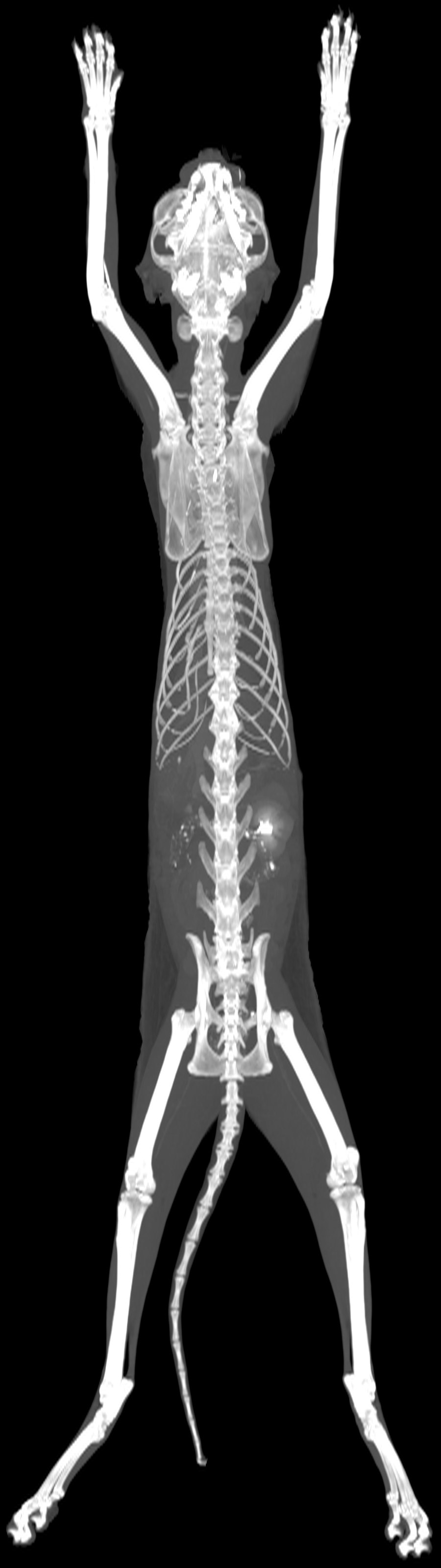
Dorsal MIP of the entire volume, post-contrast.
History
Cachexia, chronic haematemesis. Under treatment for hyperthyroidism.
Technique
4,056 slices (1 and 2 mm), pre- and post-contrast series reconstructed with medium- and high-frequency algorithms. Good study quality.
Findings
–Neck: oval, irregular mass of heterogeneous soft tissue attenuation, mildly hypoattenuating relative to the adjacent musculature, heterogeneously enhancing, located ventrally on the right, without vascular invasion or invasion of the surrounding tissues; corresponds to the right thyroid lobe (40.6 × 14.7 × 18.5 mm). Left lobe not identified.
–Thorax: enlarged sternal lymph nodes without a visible hilum. Left atrial dilatation; thickened left ventricular free wall and interventricular septum.
–Liver: hypoattenuating, non-enhancing nodular lesion in the right lateral lobe (5.5 × 7.3 × 7.8 mm); small lesion in the left medial lobe, isoattenuating on pre-contrast and hypoattenuating on post-contrast images (4.3 × 5 × 5.2 mm).
–Spleen: heterogeneous enhancement of the cranial margin; multiple irregularly shaped hypoattenuating foci.
–Kidneys: right kidney small and non-enhancing, with a triangular hyperattenuating focus at the level of the renal pelvis; left kidney enlarged, with irregular contours and heterogeneous enhancement, containing multiple oval to spherical hypoattenuating foci, some protruding beyond the renal contour.
–Stomach: diffuse circumferential wall thickening at the cardia and along the greater curvature, with homogeneous enhancement.
–Small intestine difficult to assess; multifocal fat stranding and adhesions between intestinal loops.
–Lymph nodes: enlarged gastric, splenic, cranial and caudal mesenteric, lumbar aortic, internal and medial iliac nodes, without a visible hilum.
–Peritoneum: fat stranding in the mesogastric and hypogastric regions; multifocal small volumes of free fluid.
Right-sided mandibular and parotid sialoadenopathy, multifocal pneumopathy and gastrogastric intussusception in a cat
Patient
cat, British Shorthair, neutered male, 11 years
Examination
Whole-body CT, pre- and post-contrast
Date
March 2026
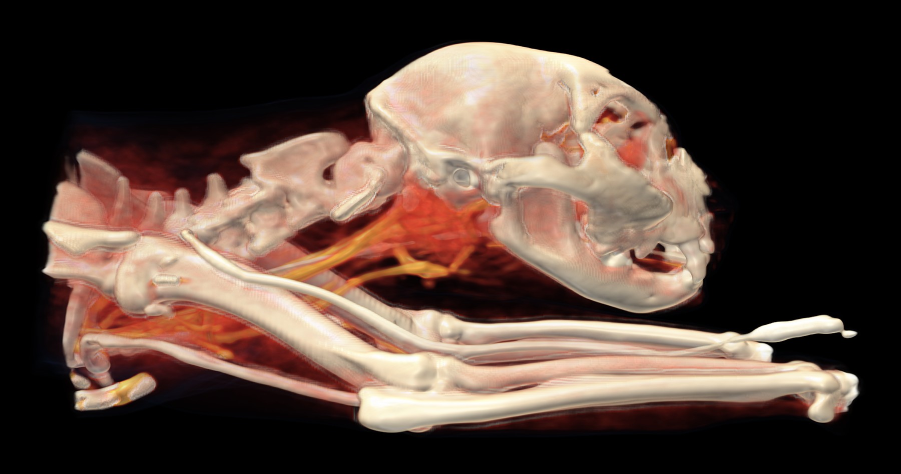
Volume rendering (VR) with an HU-based colour map from the 2 mm post-contrast series, resampled to an isotropic voxel size of 0.4 mm, right lateral view: region of right-sided sialoadenopathy and cervical lymphadenopathy, cervical vessels; along the thoracic limb, the cannula extension line containing contrast medium.
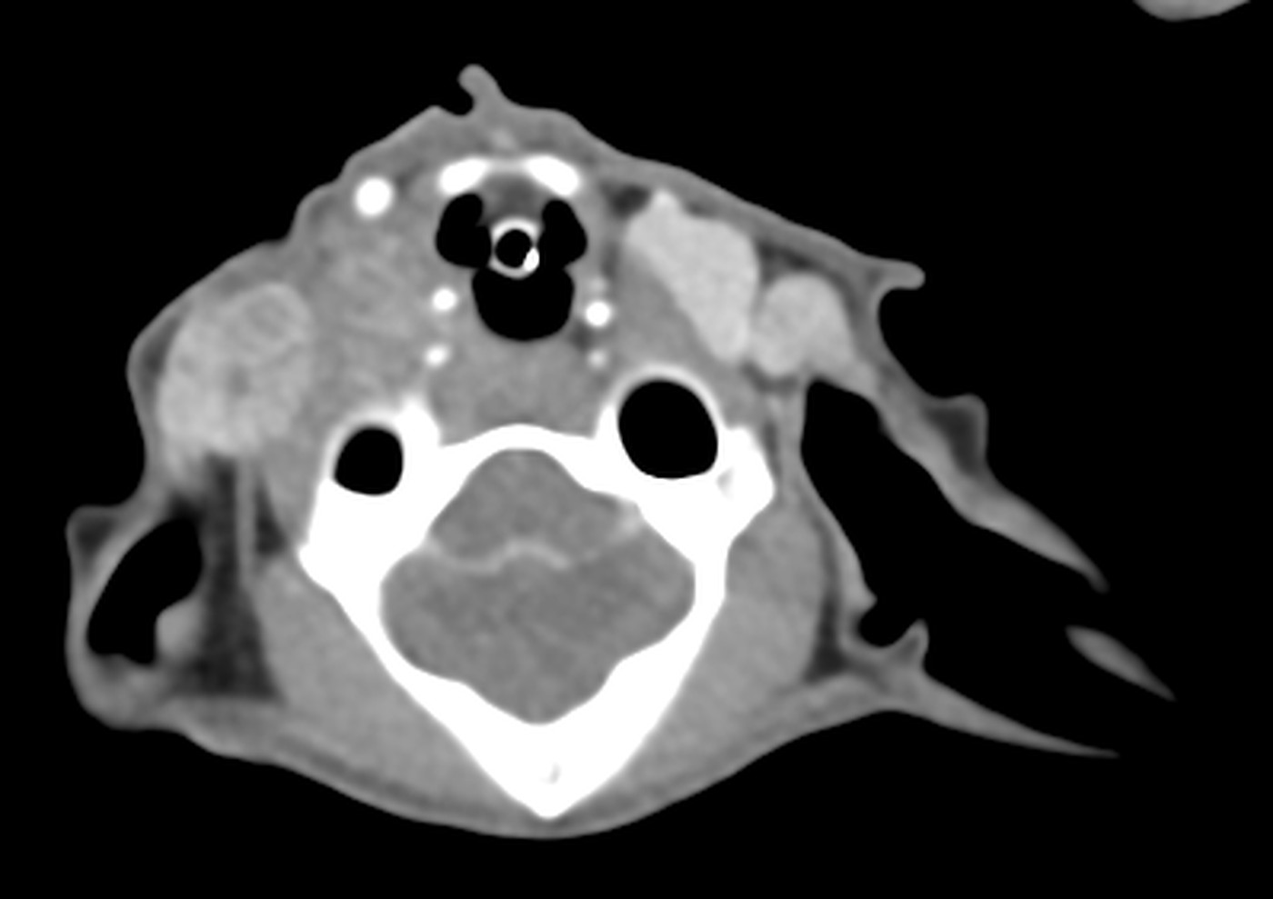
Transverse slice at the level of the skull base, post-contrast, W 400 / L 40: region of right-sided mandibular and parotid sialoadenopathy.
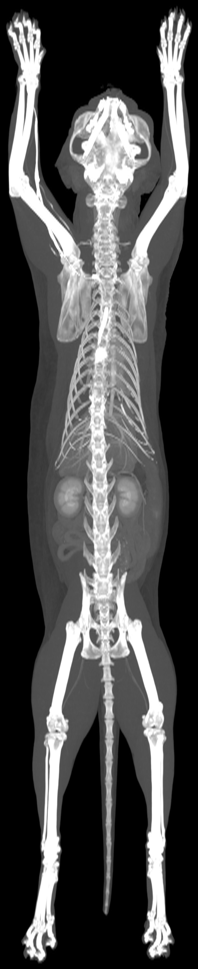
Dorsal MIP of the entire volume, post-contrast.
History
Firm mass on the neck for approximately 2 months. Pruritus of the neck and head, self-trauma.
Technique
3,811 slices (1 and 2 mm), pre- and post-contrast series reconstructed with medium- and high-frequency algorithms. Good study quality.
Findings
–Salivary glands: right mandibular and parotid glands diffusely enlarged, irregular in shape, of soft tissue attenuation, heterogeneously enhancing and poorly demarcated from the surrounding tissues.
–Lymph nodes of the head and neck: enlarged right medial retropharyngeal (spherical, heterogeneously enhancing), right parotid, bilateral ventral superficial cervical and left dorsal superficial cervical nodes, without a visible hilum.
–Lungs: small and large irregularly shaped nodules of soft tissue attenuation in all lobes, the large ones heterogeneously enhancing; multifocal ground-glass opacities, both central and subpleural; parenchymal bands.
–Heart: thickened left ventricular free wall and interventricular septum; left atrial dilatation.
–Stomach: small and spherical; cardia and fundus displaced aborally into the lumen of the gastric body, with concentric wall layering and interposed fat (intussusception); diffuse wall thickening of the gastric body (intussuscipiens); pylorus in a normal position; no mural mass or nodule.
Conclusion
–Right mandibular and parotid sialoadenopathy. DDx: sialoadenitis, neoplasia.
Biopsy and correlation with cytology or histopathology.
Note
Gastrogastric intussusception may be transient, with spontaneous resolution. The thickening of the outer wall layer (intussuscipiens) is probably a consequence of it, but may also reflect an inflammatory or neoplastic process. Gastric ultrasound is recommended.
Translated from the Czech original, with identifying details removed. Report signed by MVDr. Ladislav Stehlík, Ph.D..
Nodular lesion of the right cerebral hemisphere with destruction of the cribriform plate in a dog with epileptiform seizures
Patient
dog, Staffordshire Bull Terrier, male, 11 years
Examination
CT of the head, pre- and post-contrast
Date
August 2026
History
Epileptiform seizures and circling to the right over the past month.
Technique
1,202 slices (1 and 2 mm), pre- and post-contrast series reconstructed with medium- and high-frequency algorithms. Excellent study quality.
Findings
–CNS: heterogeneous pre-contrast attenuation of the rostral part of the right hemisphere and the olfactory lobe; heterogeneous, predominantly peripheral enhancement in the central part of the right hemisphere (23 × 17 × 12 mm); focal displacement of the falx cerebri to the left; asymmetry of the lateral ventricles (left > right), with contents of fluid attenuation; focal destruction of the right cribriform plate without extension into the nasal cavity.
–Teeth: multifocal destruction/resorption at the level of the necks of teeth 105 and 106; focal osteolysis of the alveolar bone of tooth 106; no communication with the nasal cavity.
Conclusion
–Nodular lesion in the right cerebral hemisphere. DDx: neoplasia (suspected glioma), abscess.
–Destruction of the right cribriform plate, suspected to be secondary to the brain lesion (inflammatory or neoplastic activity), or possibly pressure necrosis of the bone (oedema, raised intracranial pressure).
Recommendation
Neurological examination, MRI of the brain and CSF analysis.
Translated from the Czech original, with identifying details removed. Report signed by MVDr. Ladislav Stehlík, Ph.D..