Skip to content

We send your patient back with the report

Refer a patient today, no contract needed. You get a radiologist's report, within 24 hours on the fast track, and DICOM data to download. The client goes back to you; we do not take them over.

Partner areanext available: Friday 2 October, 08:15
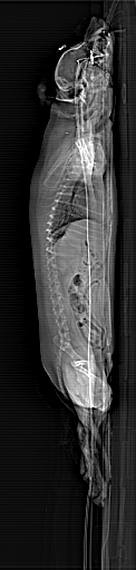
Topogram, the patient's planning scan
Topogram with the scan range marked. This is exactly how the area to be scanned is marked before each scan.

The patient is yours.
The scan is ours.

We don't take over your clients

We do not book your client in for check-ups, we do not offer them our services beyond what you referred them for, and we do not open a record for them for further treatment.

Your question sets the protocol

The report answers what you asked. If we find something else as well, we include it, but the main answer is always to your question.

It's your data

You can download the DICOM data and the report without limits: today, a year from now, or when you want a second opinion elsewhere. We do not charge for access to your patient's images.

We report findings to you

If the scan shows a condition that needs intervention, you hear it from us first. Where you send the patient next is your decision, even if it is not to us.

Two ways to refer

The quality of the examination is the same in both. What differs is who pays, who interprets the images and how much paperwork is needed at the start.

Direct referralno contractContract referralframework agreement
When you can startToday, just call.Within two working days of agreeing terms.
PricePublic rate, the same for you and the client.Rate agreed in the framework agreement.
Who paysThe client pays us directly.The client pays you, and we invoice your practice.
Who reportsAlways a vetCT radiologist.vetCT or you, depending on the agreement and the case.
Attending the scanBy arrangement.Any time, with the client too.
Partner areaNot needed.Calendar, reports, DICOM, patient overview.
PaperworkNone.Once, at the start.

What your report
will look like

A real report from our archive: CT of the neck, thorax, abdomen and pelvis, pre- and post-contrast, female patient, 11 years, August 2026. Identifying details have been removed and the report has been translated from the original Czech; the content of the findings is unchanged.

Clinical information
Dyspnoea, lethargy, anaemia.
Region examined
Neck, thorax, abdomen, pelvis
Technical parameters
4,010 slices (1 and 2 mm) available: pre- and post-contrast CT images reconstructed with medium- and high-frequency algorithms.Study of diagnostic quality. 3 post-contrast series submitted, corresponding to the late venous and excretory phases. Motion artefacts.
Findings
Full findings, from neck to spine
NECKAbnormal left superficial cervical lymph node: normal shape, enlarged, hilum absent, homogeneous attenuation, homogeneous enhancement.THORAXTRACHEA AND BRONCHINormal findings.LUNGSMultiple miliary and small nodules of soft tissue attenuation, diffusely throughout the lungs.Right cranial lobe, cranial part of the left cranial lobe: consolidation of the ventral third of the lung parenchyma, air bronchograms.Remaining lung lobes: ground-glass opacity to consolidation with air bronchograms ventrally, and in the right middle lobe also in the central third, diffuse distribution.PLEURANormal pleural space.OESOPHAGUSNormal oesophageal wall and lumen.LYMPH NODESAbnormal left axillary lymph nodes: normal shape, mildly enlarged, hilum absent, homogeneous attenuation, homogeneous enhancement.Abnormal sternal lymph nodes: irregular shape, enlarged, hilum absent, homogeneous attenuation, homogeneous enhancement.One abnormal cranial mediastinal lymph node: normal shape, mildly enlarged, hilum absent, homogeneous attenuation, heterogeneous enhancement.Abnormal tracheobronchial lymph nodes: irregular shape, enlarged, hilum absent, homogeneous attenuation, heterogeneous enhancement.CARDIOVASCULAR SYSTEMNormal.THYMUSNormal.MEDIASTINUMNormal.THORACIC WALL AND DIAPHRAGMNormal findings.ABDOMENLIVER AND BILIARY SYSTEMDiffusely enlarged liver, soft tissue attenuation, homogeneous enhancement, rounded and smooth margins.Gallbladder wall thickened and homogeneously enhancing, with a peripheral rim of fluid attenuation (sandwich appearance), small volume, normal position, mineralised sediment. Common bile duct normal.SPLEENDiffusely enlarged, rounded and smooth margins, normal attenuation, homogeneous enhancement.PANCREASNormal findings.ADRENAL GLANDSNormal findings.URINARY SYSTEMNormal findings.GASTROINTESTINAL TRACTNormal findings.REPRODUCTIVE SYSTEMConsistent with neutering.VASCULAR SYSTEMNormal findings.LYMPH NODESAbnormal hepatic lymph nodes: spherical shape, enlarged, hilum absent, homogeneous attenuation, heterogeneous enhancement.Abnormal splenic lymph nodes: irregular shape, enlarged, hilum absent, homogeneous attenuation, heterogeneous enhancement.Abnormal lumbar aortic and medial iliac lymph nodes: normal shape, mildly enlarged, hilum absent, homogeneous attenuation, heterogeneous enhancement.PERITONEUM/RETROPERITONEUMMultifocal free fluid in the peritoneal cavity.ABDOMINAL WALL AND PERINEAL REGIONNormal findings.PELVISNormal findings.THORACIC LIMBSNormal findings within the field of view.PELVIC LIMBSBilateral osteoproliferation of the femoral heads and acetabula.SPINEMultiple osteoproliferations of the lumbar vertebral bodies, not extending into the neuroforamina or the vertebral canal.Osteoproliferation of the L7-S1 vertebral bodies and narrowing of the right L7-S1 neuroforamen.
Conclusion
Diffuse hepatomegaly and splenomegaly. Multifocal lymphadenopathy. DDx: lymphoma, systemic infections, extramedullary haematopoiesis (splenomegaly alone may be secondary to anaemia).Gallbladder wall thickening consistent with oedema. DDx: anaphylaxis, cholecystitis, hypoalbuminaemia.Ascites, small volume. DDx: neoplastic, secondary to hepatopathy.Pulmonary consolidation with this distribution is most consistent with pneumonia.Pulmonary ground-glass opacity. DDx: lymphoma, interstitial pneumopathy, pneumonia. These may also result from motion artefacts and/or anaesthesia.INCIDENTAL FINDINGSSpondylosis deformans.DLSS
Recommendations and notes
For further assessment, I recommend biopsy and correlation with cytology or histopathology.

Report signed by MVDr. Ladislav Stehlík, Ph.D.
Case studies from our archive

The partner area
requires verification

Partner area

Access for partner practices

Enter the email address or phone number we hold for your practice. We will send a four-digit code to it, just as the hospital chat does when it recognises you. There is no password to remember.

  • Booking patients at your partner rate
  • Reports and image data for your patients, ready to download
  • An overview of everything you have referred to us

Not working with us yet? Enter your email and we will take you straight to the application form.

What we
scan most often

Other areas we scan: neck, dental CT, emergency medicine, polytrauma. We adapt the scan range to the question you need answered.

Rates for practices

Prices include anaesthesia and VAT. The lower price applies to a standard report, the higher one to a report within 24 hours. A patient over 8 years old or unwell needs a pre-anaesthetic examination before CT. We can do it here for €89, or you can upload results no older than 7 days. You will see the total in the order summary and pay it at the clinic at the examination. Contract practices have a rate agreed in their framework agreement and are invoiced once a month. Details, including cancellation, are in the terms and conditions.

One scan,
every view

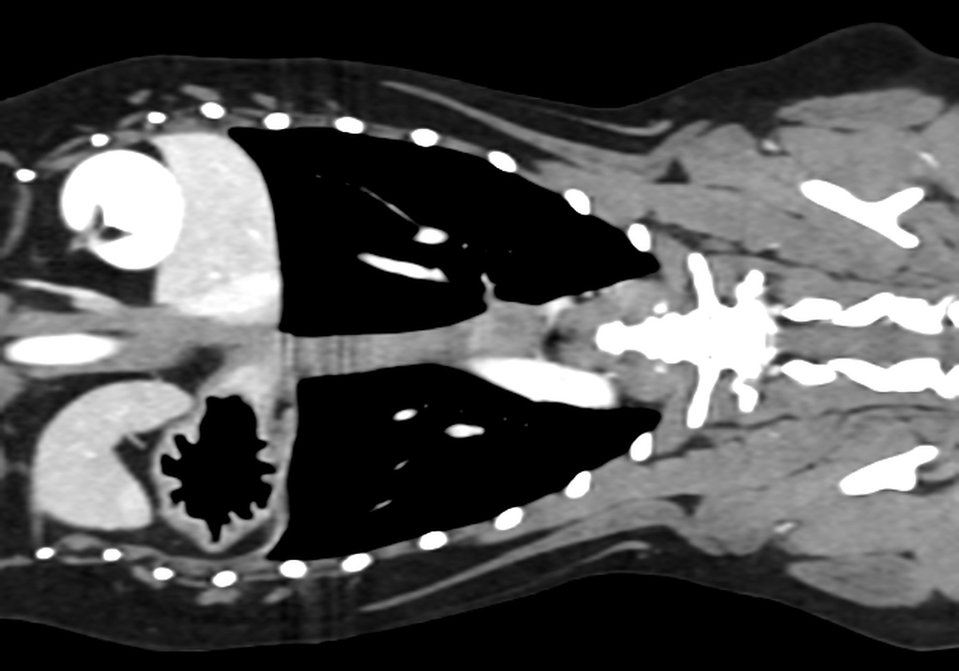
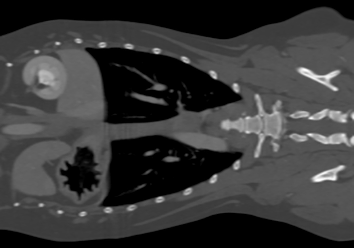
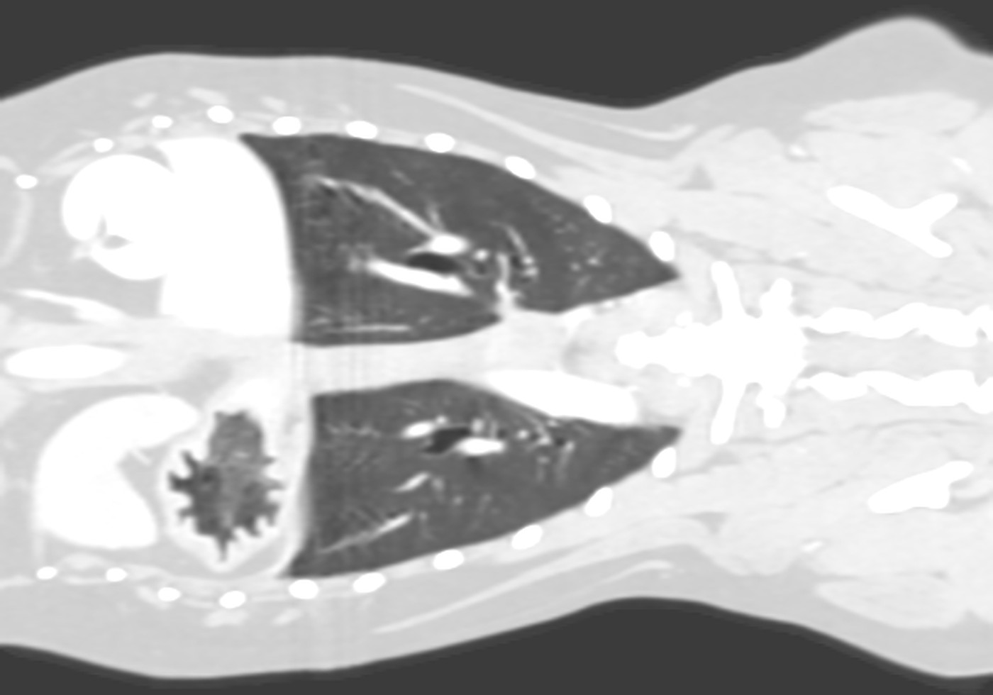
Longitudinal CT slice through a dog's chest, soft tissue windowLongitudinal CT slice through a dog's chest, bone windowLongitudinal CT slice through a dog's chest, lung windowW 400 / L 40

A scan is not a single picture. It is a volume of densities: an X-ray merges the whole thickness of the body into one grey layer, while CT assigns a density to every point in space. That is why we read the same slice three times, for organs, for bone and for lungs, and from the same volume we build a skeleton without soft tissue, or vessels without bone. With no extra radiation and no extra anaesthesia.

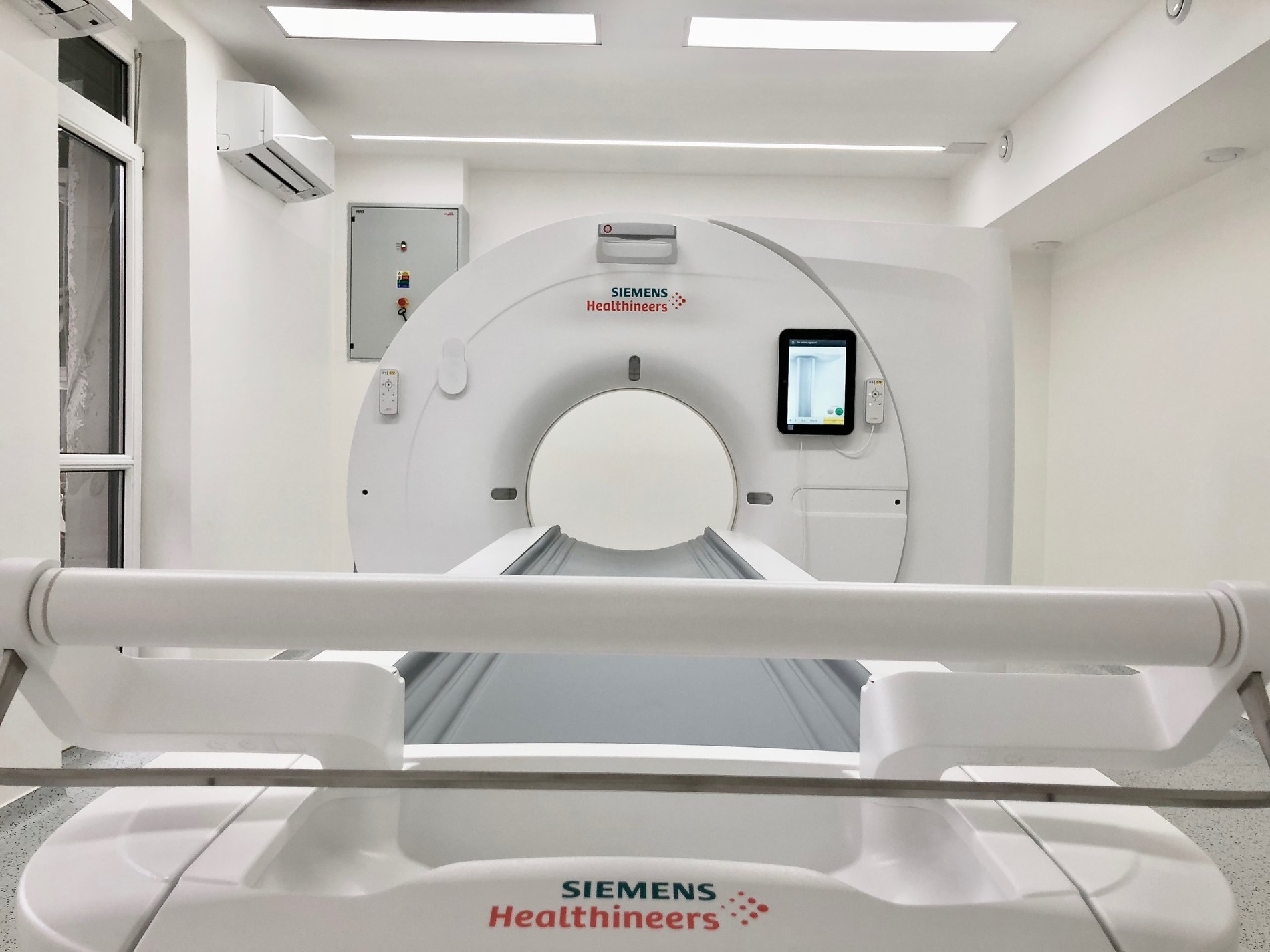
We scan on a Siemens SOMATOM go.Now with the Stellar detector: 32 submillimetre slices per rotation, SAFIRE iterative reconstruction and automatic dose adjustment to the size of the patient.

Slices per rotation
32
Slice thickness
0.6 to 2.0 mm
Scan time
under 20 seconds
Dose reduction (SAFIRE)
up to 60%
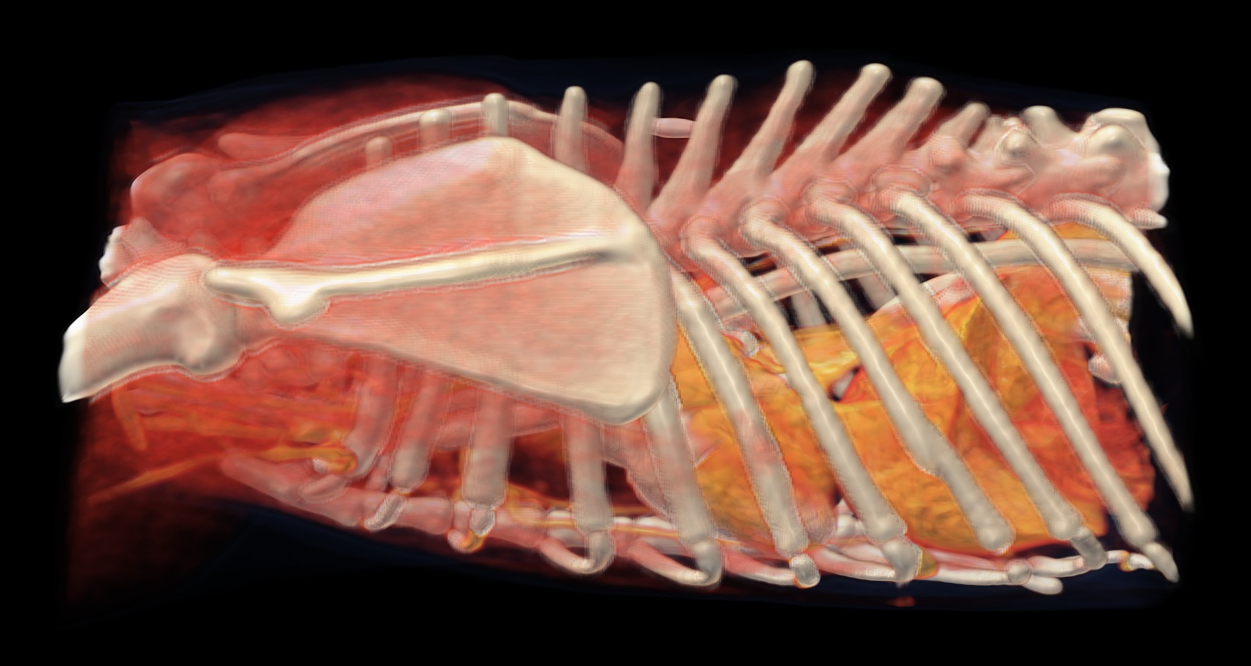
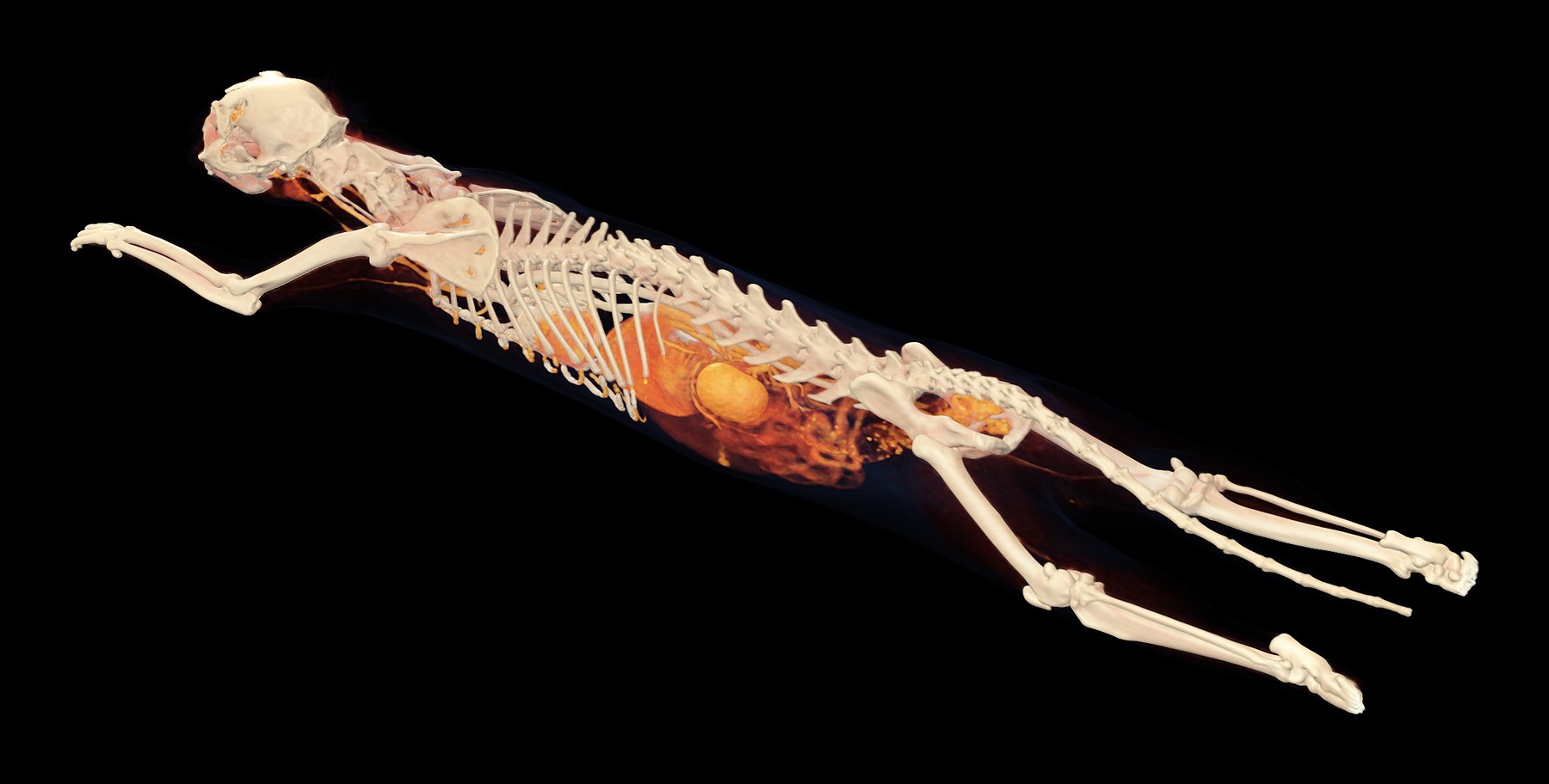
3D reconstruction of a cat's whole body from CT
The whole skeleton from a single scan. Muscle and fat are muted, so the surgeon can see the fracture and the shape of the bone before reaching for the scalpel.
Scanning room with the Siemens SOMATOM go.Now
The scanning room on Staromestská. The patient lies on a moving table, and staff watch them through the glass and on the vital signs monitor.
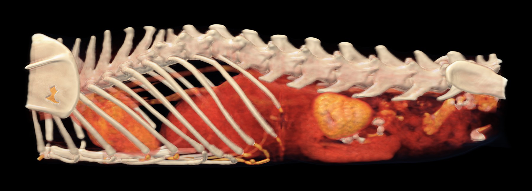
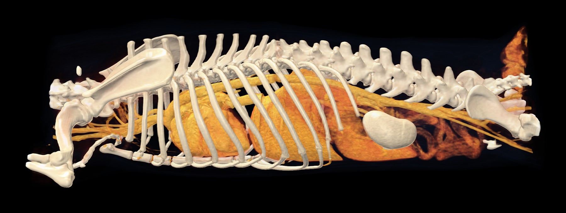
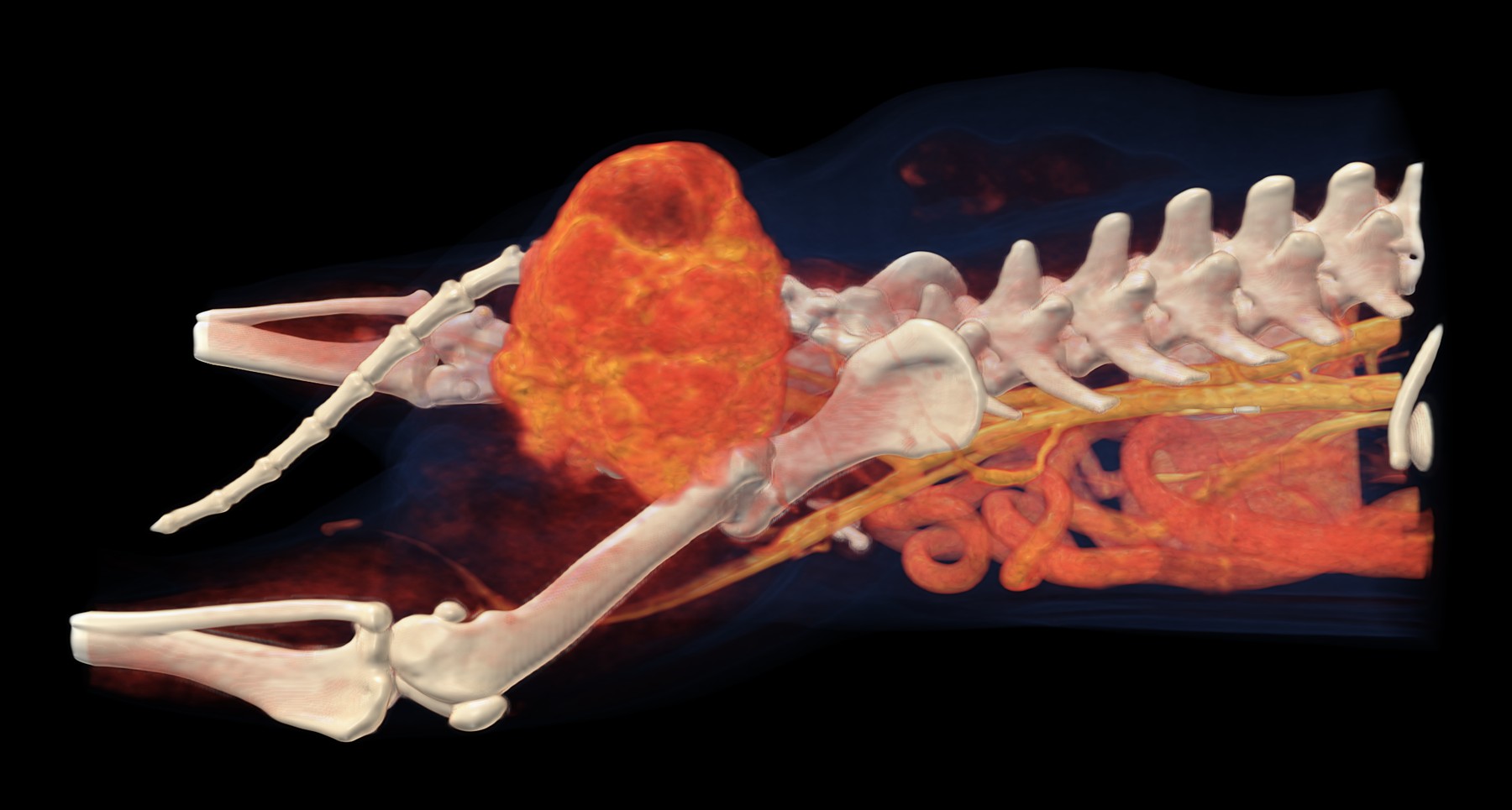
3D reconstruction of a dog's trunk with organs enhanced by contrast medium
The same scan without muscle and fat: what remains is the skeleton, and the organs and vessels enhanced by contrast medium.

A machine does the scan.
A person makes the diagnosis.

MVDr. Ladislav Stehlík, Ph.D.

Clinical lead

Academic in diagnostic imaging: radiology, ultrasonography and computed tomography. Training placements under the supervision of Prof. Massimo Vignoli, Dip. ECVDI.

MVDr. Tamara Zimanová

Reporting vet

She has worked in diagnostic imaging since graduating. At the hospital she is also responsible for digital radiography, so she knows the patients both before and after CT.

Staromestská 6/D,
Bratislava

Phone+421 2 2102 8700Emailvetct@vetline.skAddressStaromestská 6/D, 811 03 Bratislava
Scanning hours
Monday to Fridayby available appointments
Saturday and Sundayurgent cases, by arrangement

Urgent cases are seen as a priority: just call us. You can have your first appointment even without a partnership agreement; we can catch up on the paperwork afterwards.

Our CT unit is in the building of the Vetline veterinary hospital, which is open 24/7. With anaesthesia, this is no small detail: if something goes wrong, help is right next door, not a drive away.