Oncology, Abdomen, Head and neck
She was losing weight and vomiting blood. CT showed the problem was not just the thyroid
- Patient
- cat, domestic shorthair, spayed female, 16 years
- Examination
- Whole-body CT, pre- and post-contrast
- Date
- March 2026
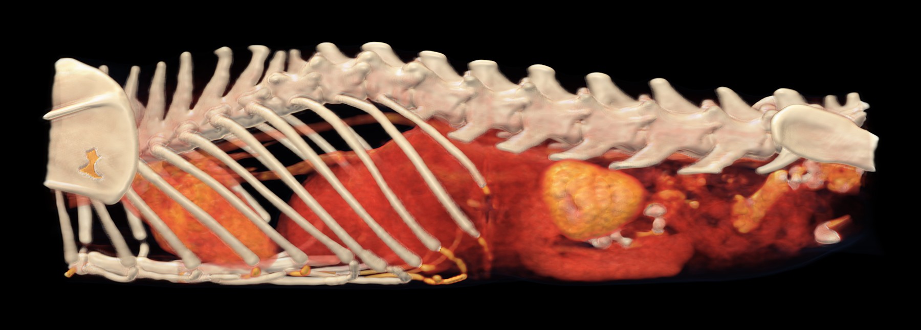
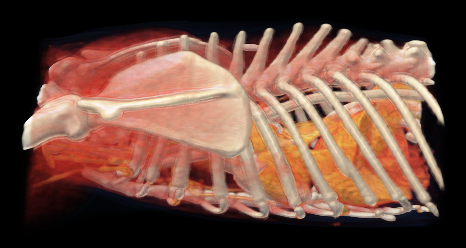
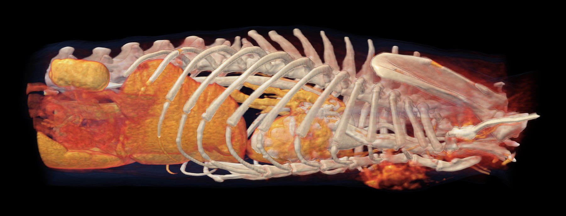
3D reconstruction of the chest and abdomen, seen from the left. Behind the ribs is the enlarged left kidney (gold), in which the CT found nodules; further forward, in the chest, is the heart.
- bone
- strongest contrast uptake: blood vessels, heart, kidneys
- organs and tissues with weaker contrast uptake, including tumours
- skin and fat
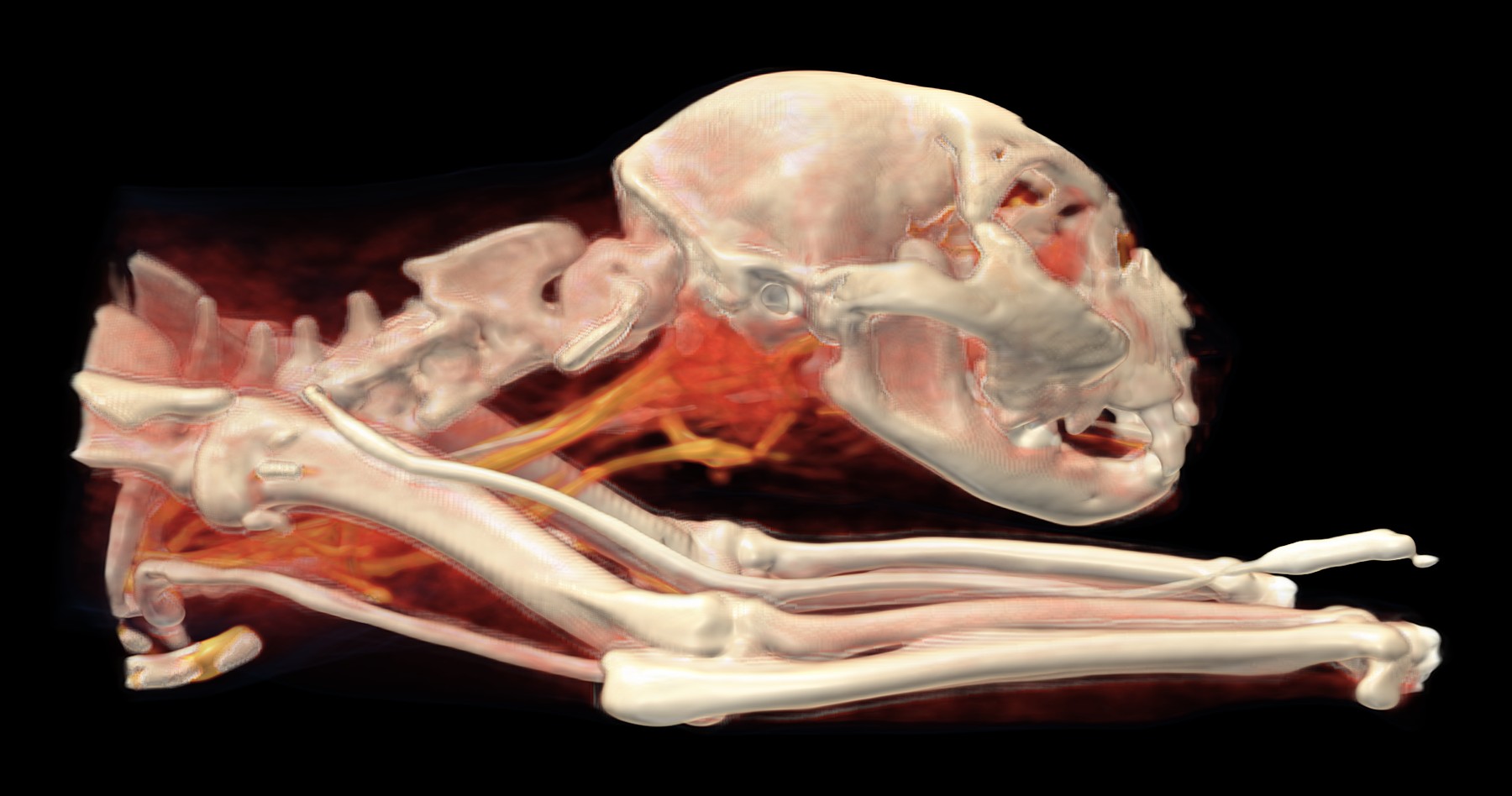
A sixteen-year-old cat was being treated for an overactive thyroid gland. Despite this, she was losing a lot of weight and had been vomiting blood for a long time. The vet needed to know what was happening throughout her body and therefore recommended a CT scan from head to tail.
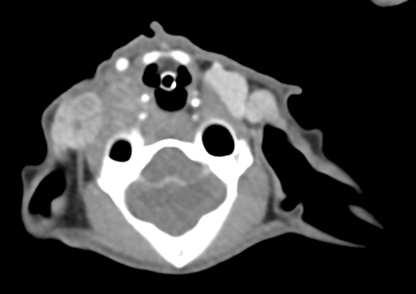
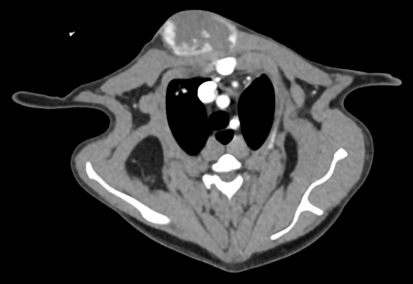
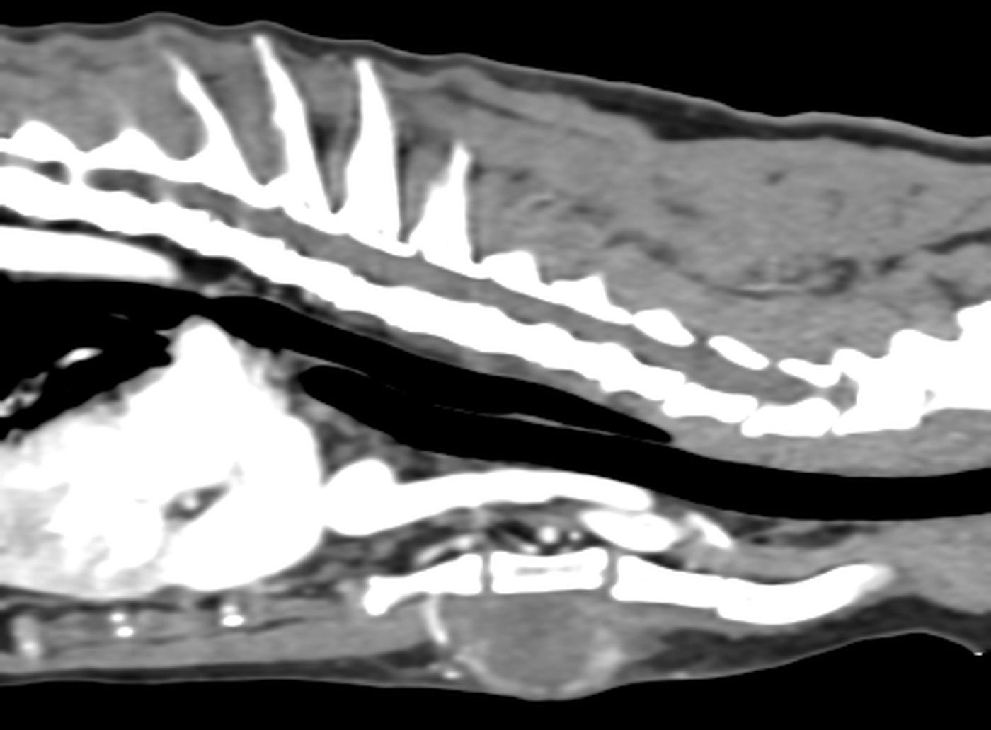
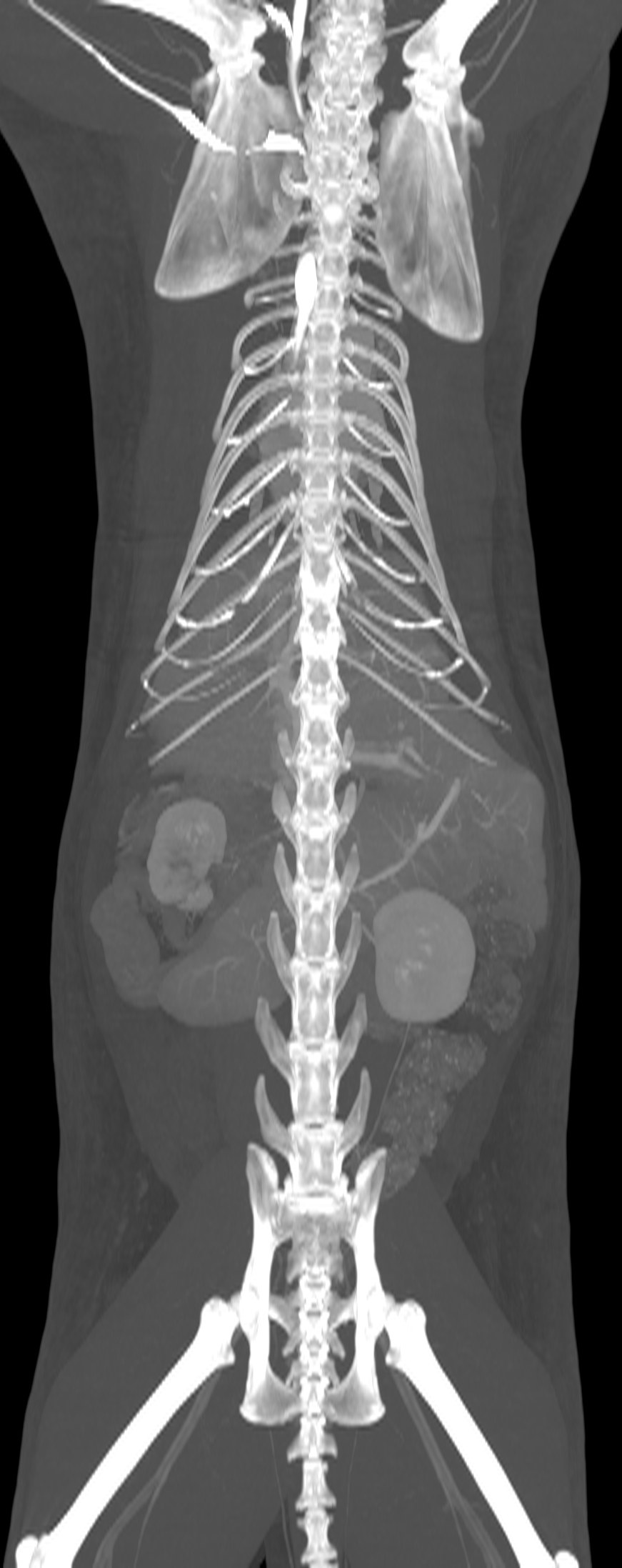
In the neck, the CT found an enlarged right lobe of the thyroid gland, a mass about four centimetres long that was not growing into the surrounding tissue. However, it also showed things that an examination of the thyroid alone would not have revealed: a thickened stomach wall, enlarged lymph nodes in several places, lesions in the spleen, tiny nodules in the liver, nodules in the left kidney and a little fluid in the abdomen. The right kidney was small, which is consistent with a long-standing disease.
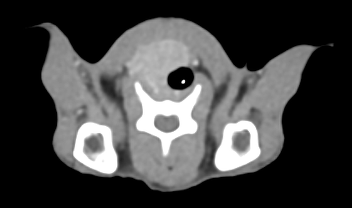
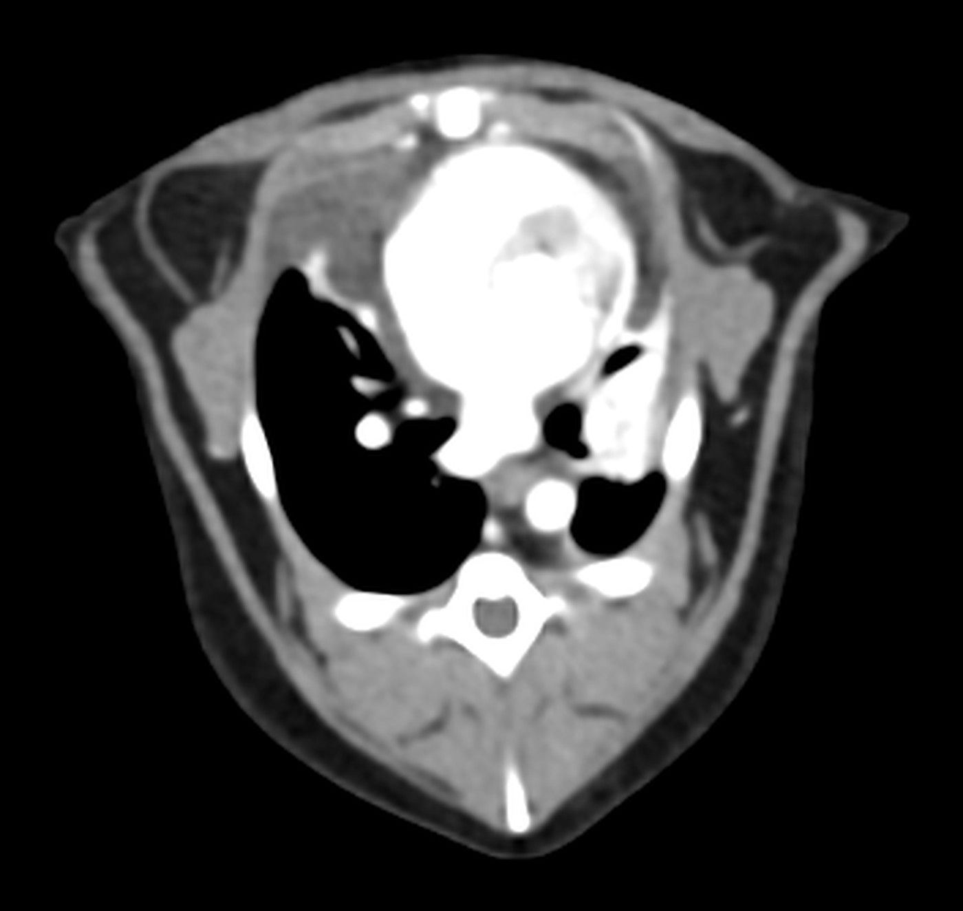
Cross-section through the neck: next to the windpipe is a non-uniform mass, the enlarged right lobe of the thyroid gland.
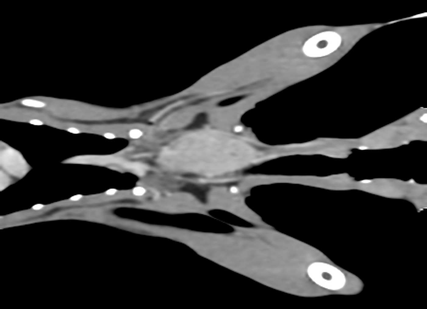
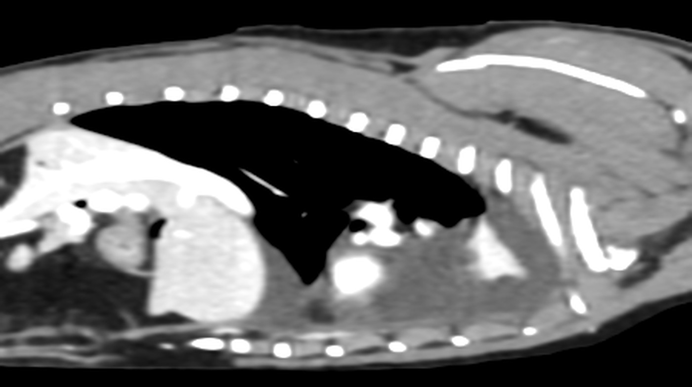
Longitudinal slice through the neck, seen from above: the oval mass in the centre is the enlarged right lobe of the thyroid gland.
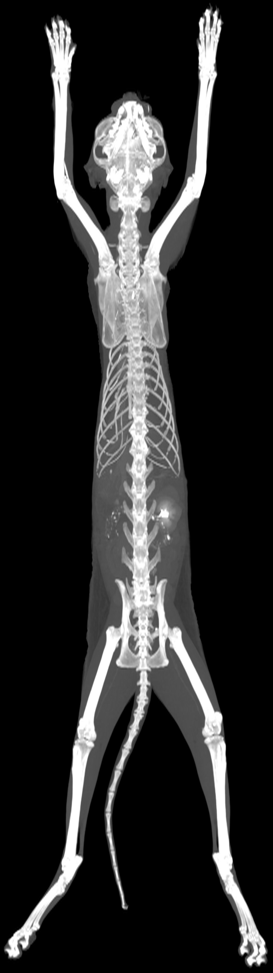
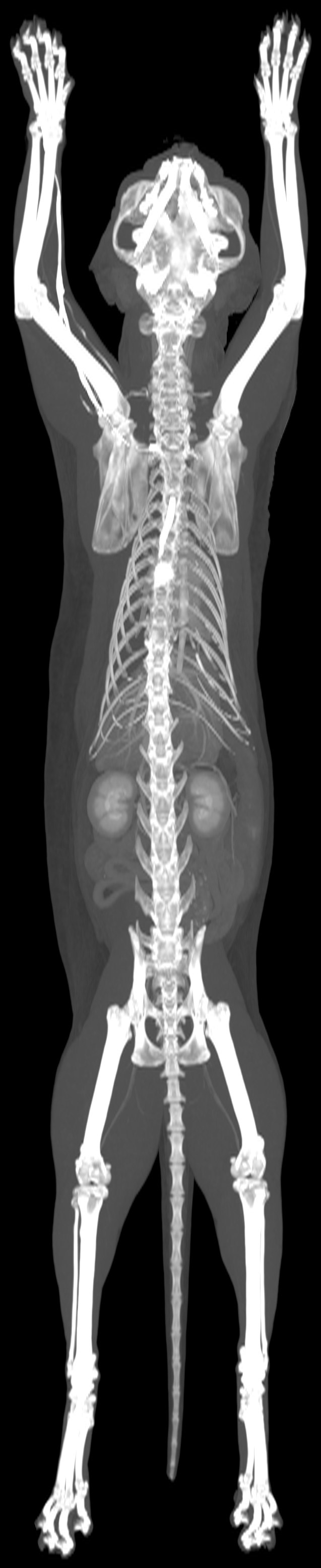
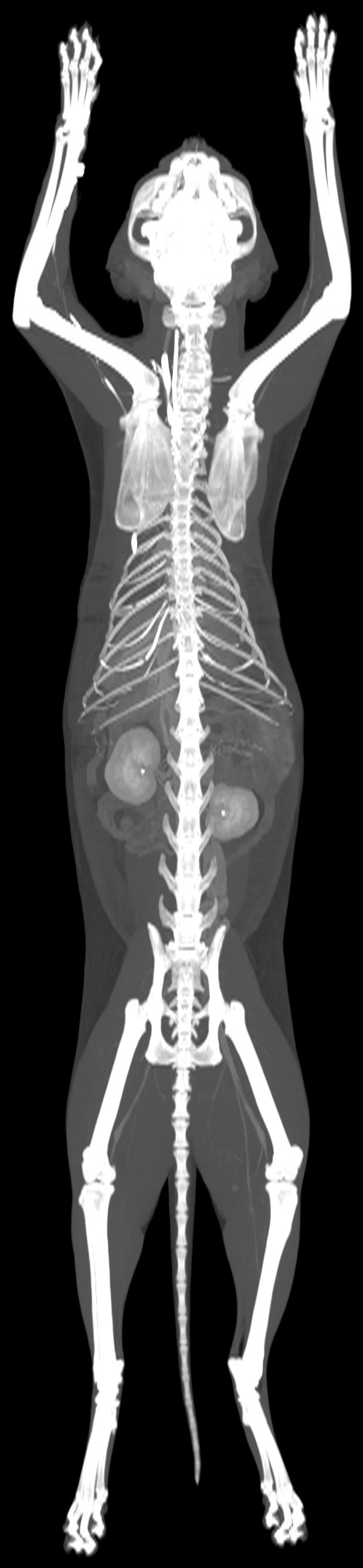
The whole scan in a single image: the skeleton, and the kidneys highlighted by the contrast agent.