Skip to content

Stories
from our CT

Real cases from our archive: what the patient came in with, why their vet recommended CT and what the scan showed. Explained so that anyone can follow, and without names.

Oncology, Abdomen, Head and neck

She was losing weight and vomiting blood. CT showed the problem was not just the thyroid

Patient
cat, domestic shorthair, spayed female, 16 years
Examination
Whole-body CT, pre- and post-contrast
Date
March 2026
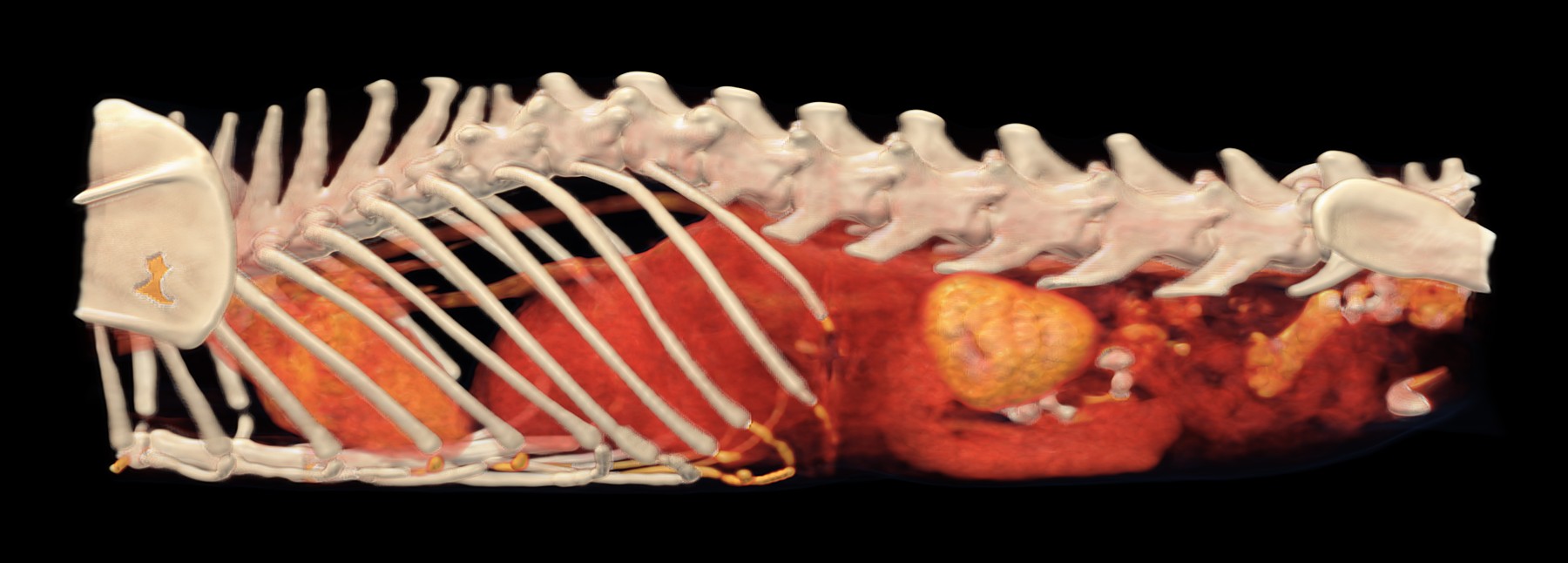
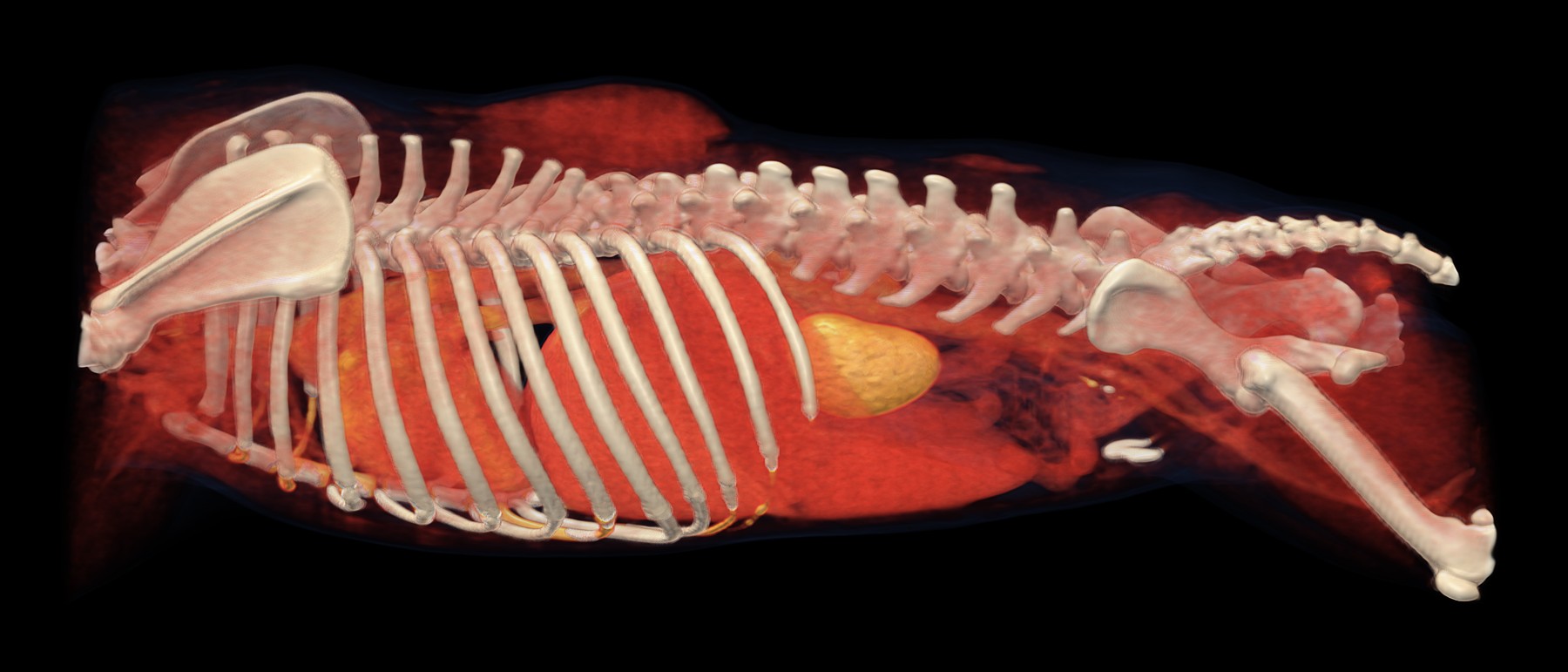
3D CT reconstruction

3D reconstruction of the chest and abdomen, seen from the left. Behind the ribs is the enlarged left kidney (gold), in which the CT found nodules; further forward, in the chest, is the heart.

  • bone
  • strongest contrast uptake: blood vessels, heart, kidneys
  • organs and tissues with weaker contrast uptake, including tumours
  • skin and fat

A sixteen-year-old cat was being treated for an overactive thyroid gland. Despite this, she was losing a lot of weight and had been vomiting blood for a long time. The vet needed to know what was happening throughout her body and therefore recommended a CT scan from head to tail.

In the neck, the CT found an enlarged right lobe of the thyroid gland, a mass about four centimetres long that was not growing into the surrounding tissue. However, it also showed things that an examination of the thyroid alone would not have revealed: a thickened stomach wall, enlarged lymph nodes in several places, lesions in the spleen, tiny nodules in the liver, nodules in the left kidney and a little fluid in the abdomen. The right kidney was small, which is consistent with a long-standing disease.
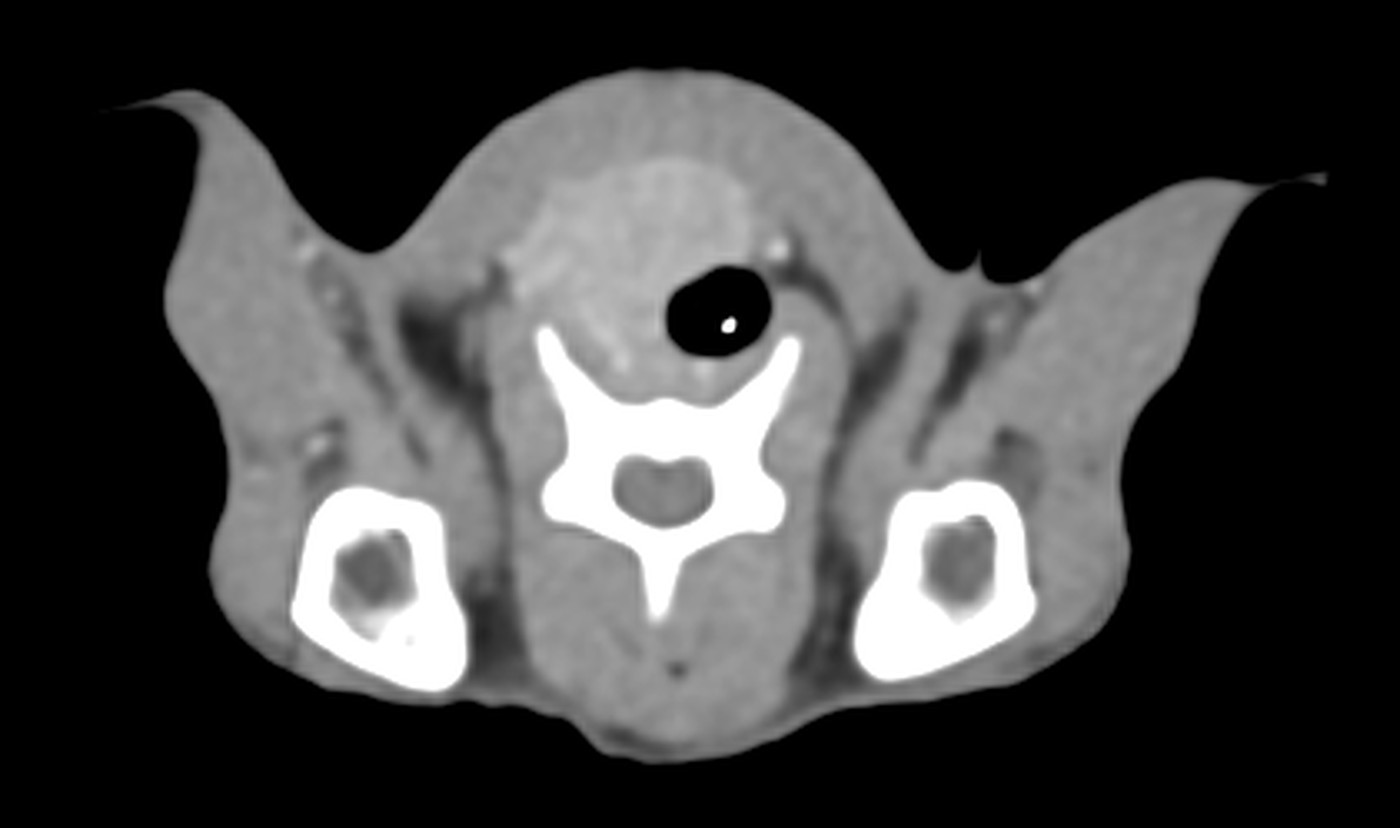
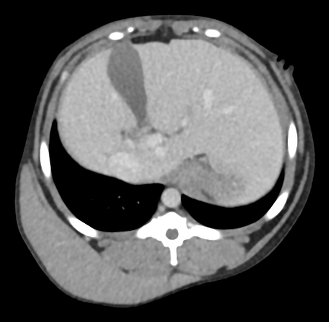
Transverse CT slice at the level of the finding

Cross-section through the neck: next to the windpipe is a non-uniform mass, the enlarged right lobe of the thyroid gland.
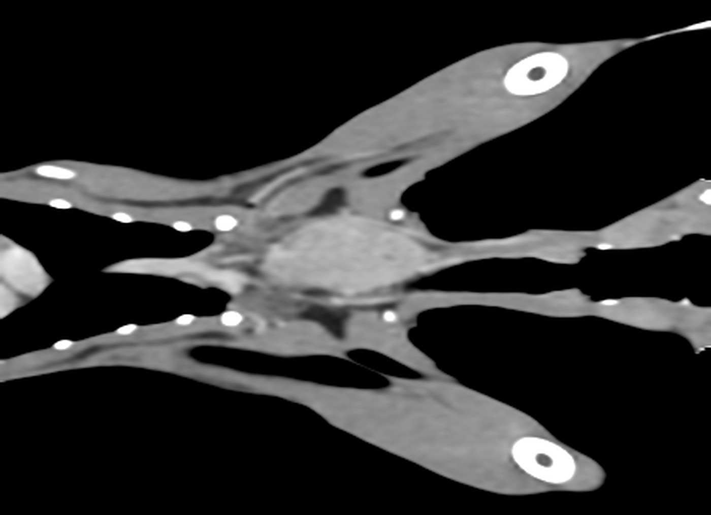
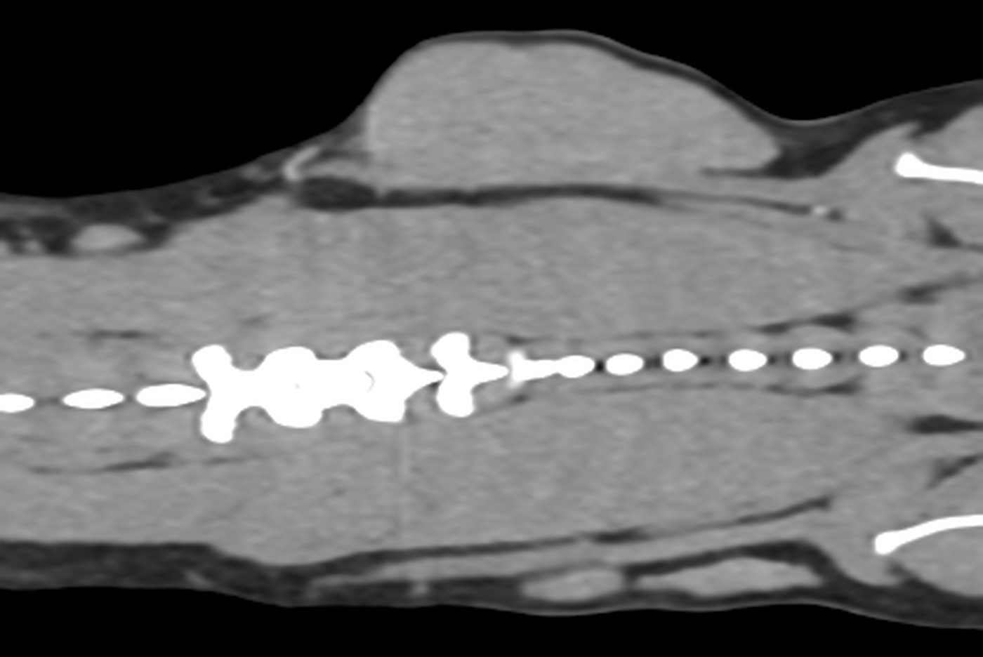
Longitudinal CT slice through the neck

Longitudinal slice through the neck, seen from above: the oval mass in the centre is the enlarged right lobe of the thyroid gland.
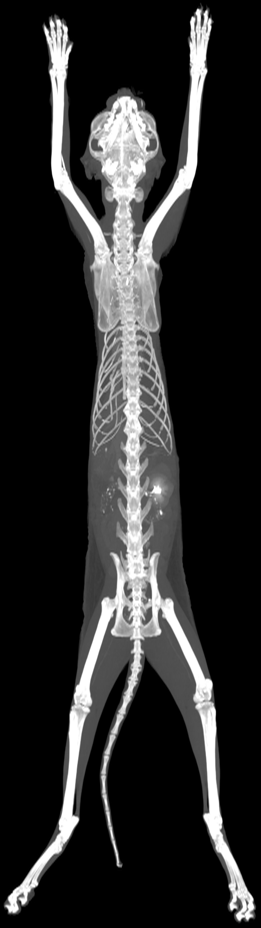
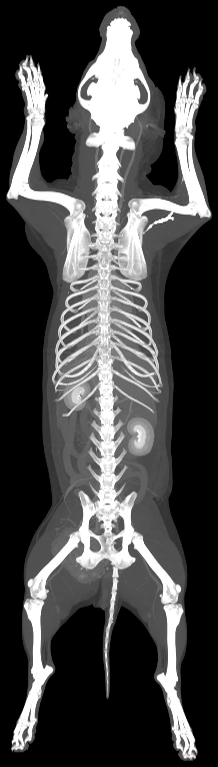
Maximum intensity projection of the whole scan

The whole scan in a single image: the skeleton, and the kidneys highlighted by the contrast agent.
Nose and ear, Chest and lungs, Oncology

He came in because of his ear. CT also found a mass near the heart

Patient
dog, French Bulldog, male, 7 years
Examination
Whole-body CT, pre- and post-contrast
Date
March 2026

A seven-year-old French Bulldog had started tilting his head to the right and had a discharge from his right ear. To find out what was going on in the ear and deeper behind it, he had a whole-body CT scan.

The CT confirmed inflammation of both outer ear canals, with a build-up of secretions on the right, and changes in the middle ear cavities that are common in flat-faced breeds. At the back of the nose it showed aberrant nasal turbinates, in other words tissue that should not normally be there.

The most important finding, however, was one the dog had not come in for: a mass about five centimetres in size at the base of the heart, wrapping around the aorta and pressing on both pulmonary arteries, and next to it fluid in the sac around the heart. An examination focused only on the ear would have left it hidden.

Head and neck, Chest and lungs, Abdomen

An itchy lump on the neck, and everything the CT showed

Patient
cat, British Shorthair, neutered male, 11 years
Examination
Whole-body CT, pre- and post-contrast
Date
March 2026
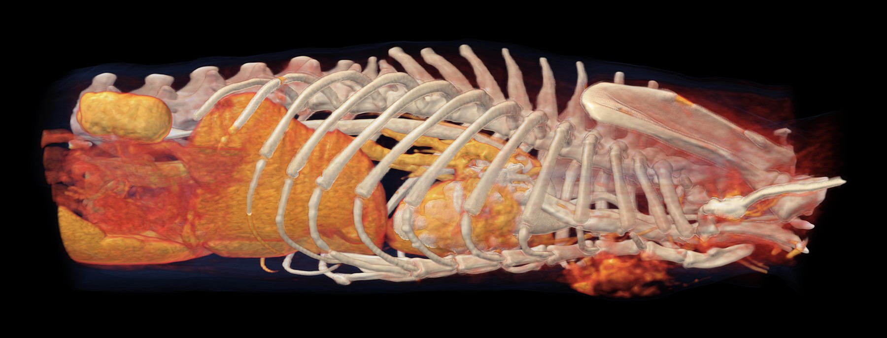
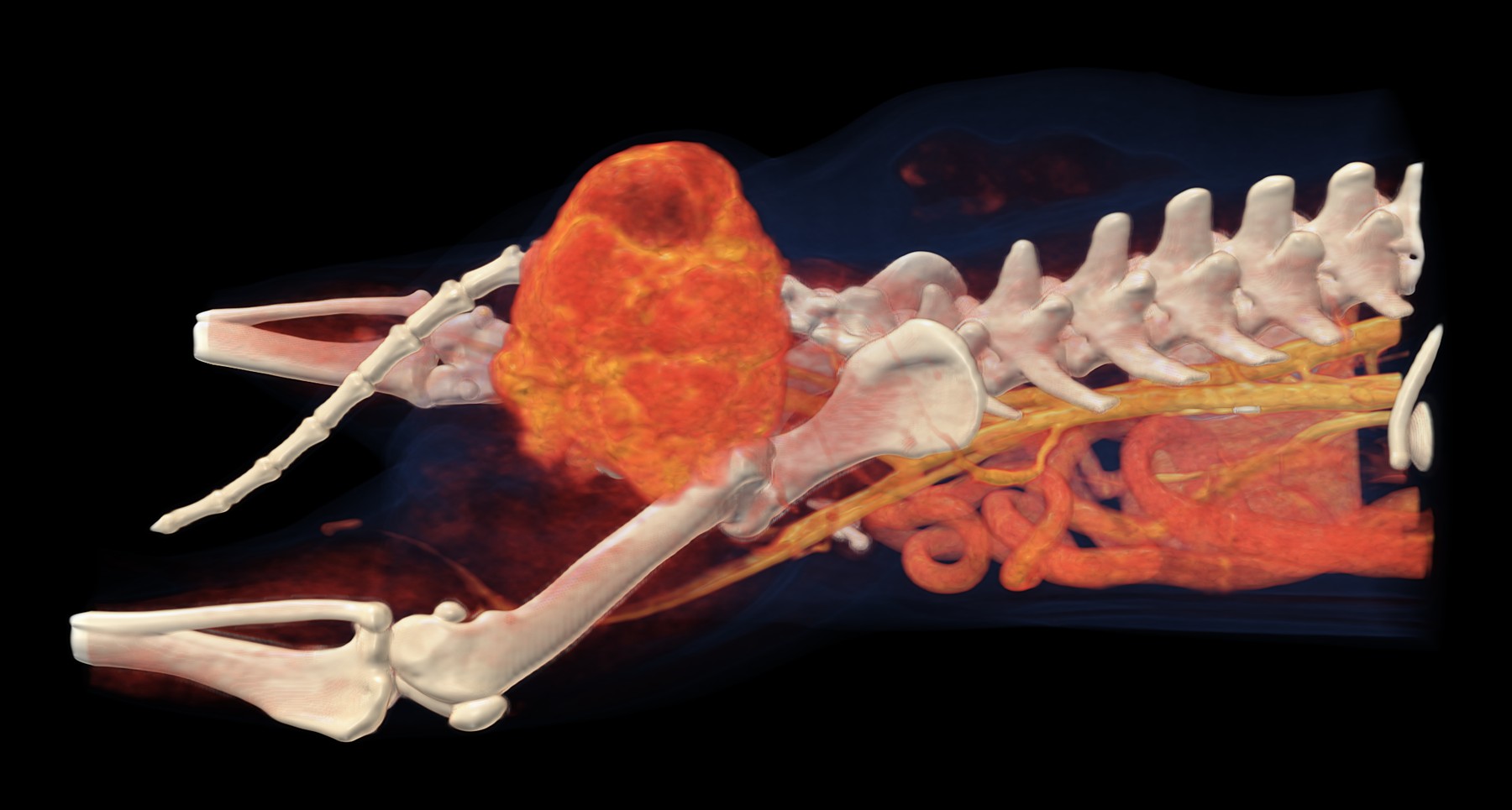
3D CT reconstruction

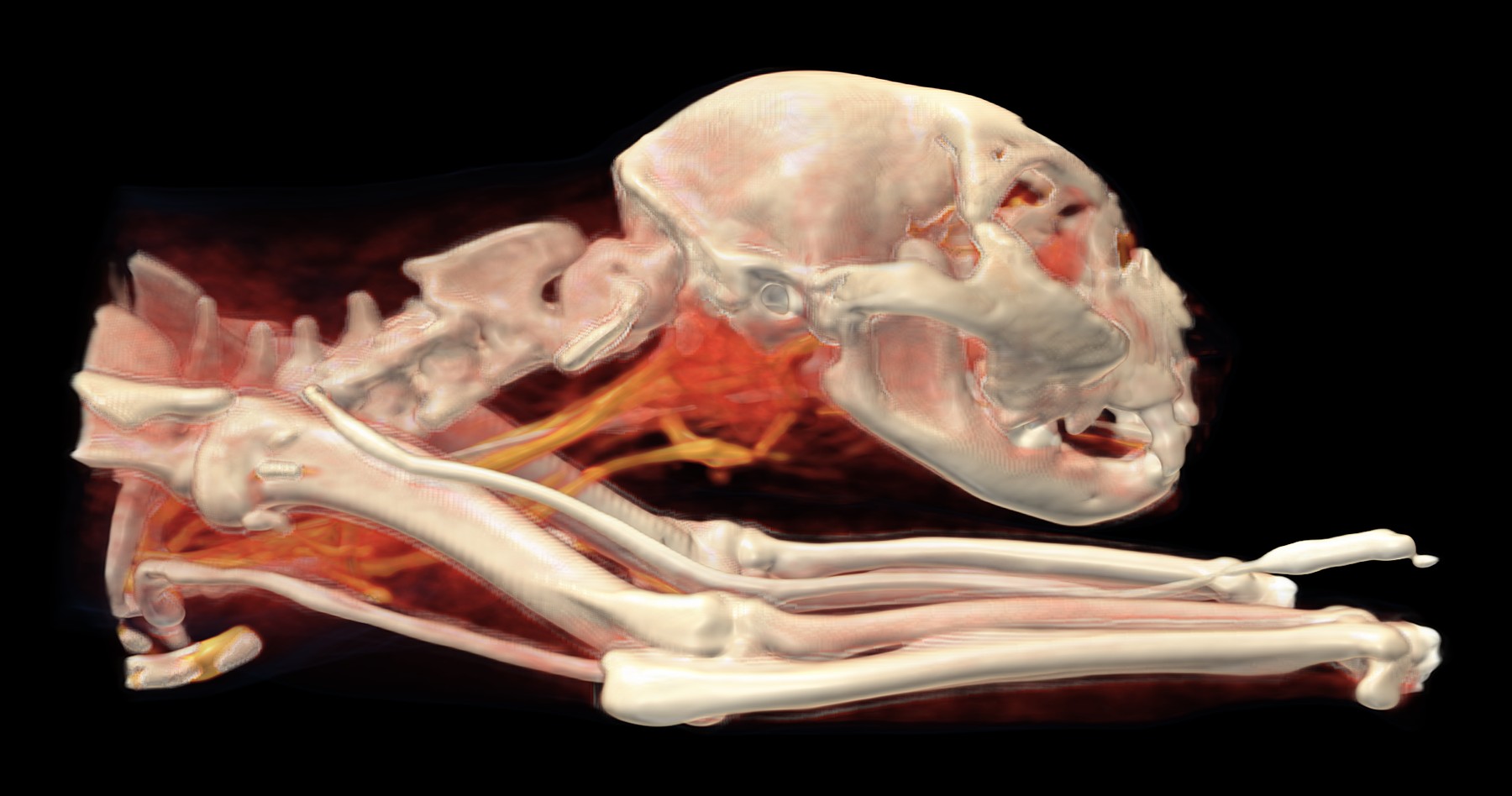
3D reconstruction of the head and neck, seen from the right. Below the skull, behind the lower jaw, is the area of enlarged salivary glands and lymph nodes (red-orange); blood vessels containing contrast agent run through the neck (gold). The white tube along the front leg is the cannula through which the contrast agent is given.

  • bone
  • strongest contrast uptake: blood vessels, heart, kidneys
  • organs and tissues with weaker contrast uptake, including tumours
  • skin and fat

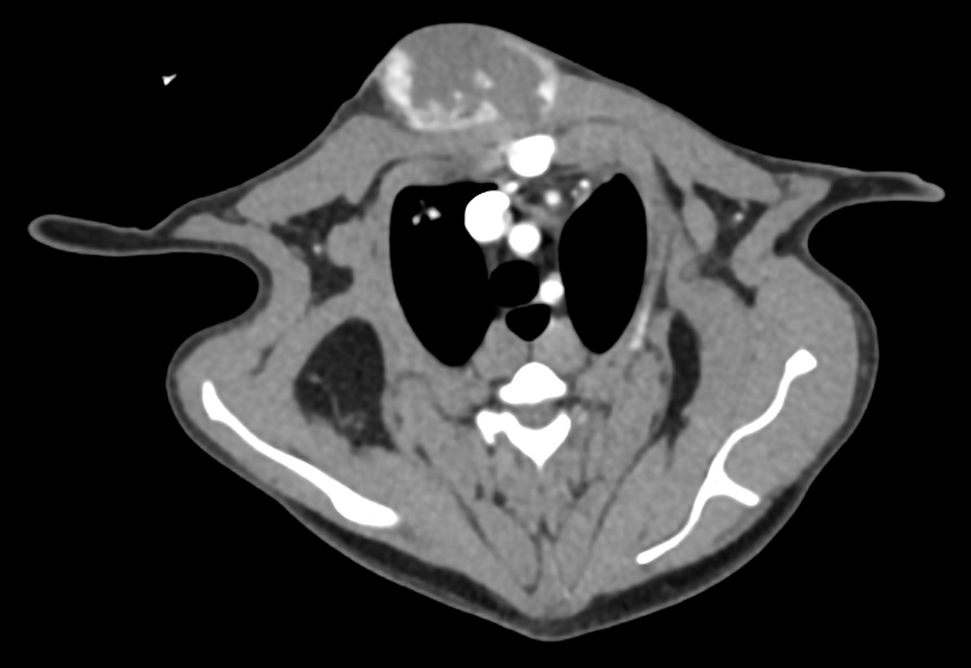
An eleven-year-old male cat had had a firm lump on his neck for about two months. His neck and head were so itchy that he was injuring himself. The vet recommended a whole-body CT scan to see where the lump was coming from and whether the problem was limited to the neck.

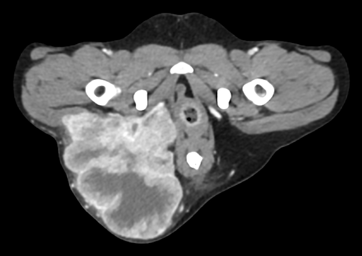
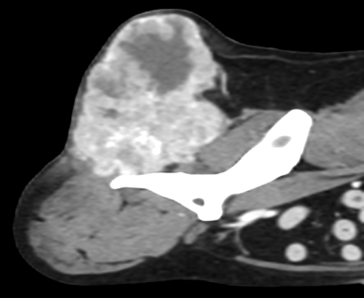
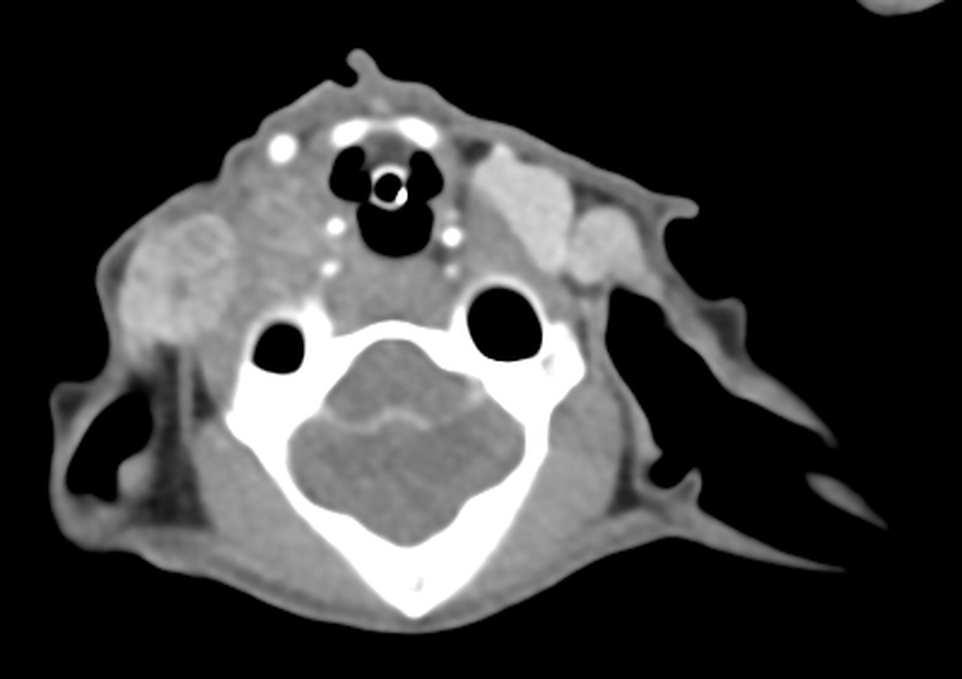
The lump turned out to be enlarged salivary glands below the right side of the lower jaw and near the ear, together with enlarged lymph nodes in the neck. However, the CT also found lesions in both lungs (small and large nodules and changes in the lung tissue) and an intussusception in the stomach, a condition in which one part of the stomach slides into another, like the sections of a telescope.

Transverse CT slice at the level of the finding

Cross-section through the upper part of the neck at the level of the abnormal salivary glands on the right.

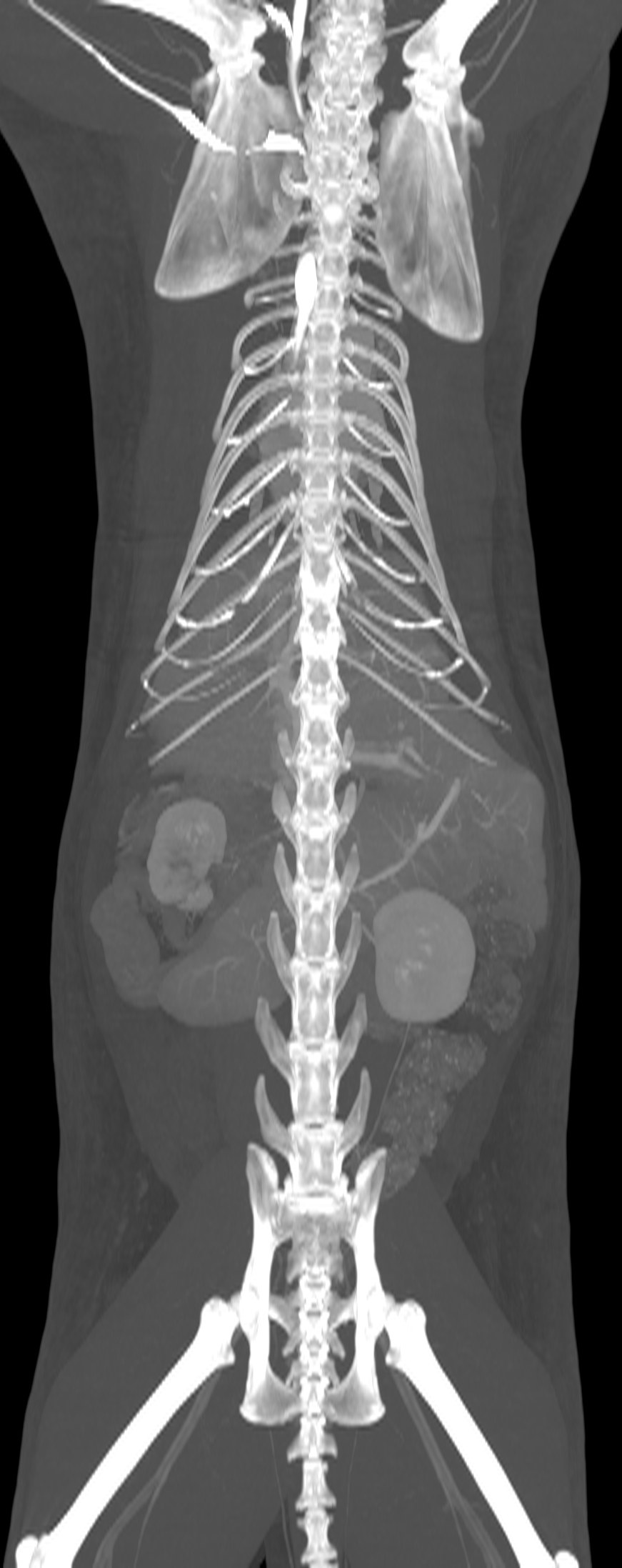
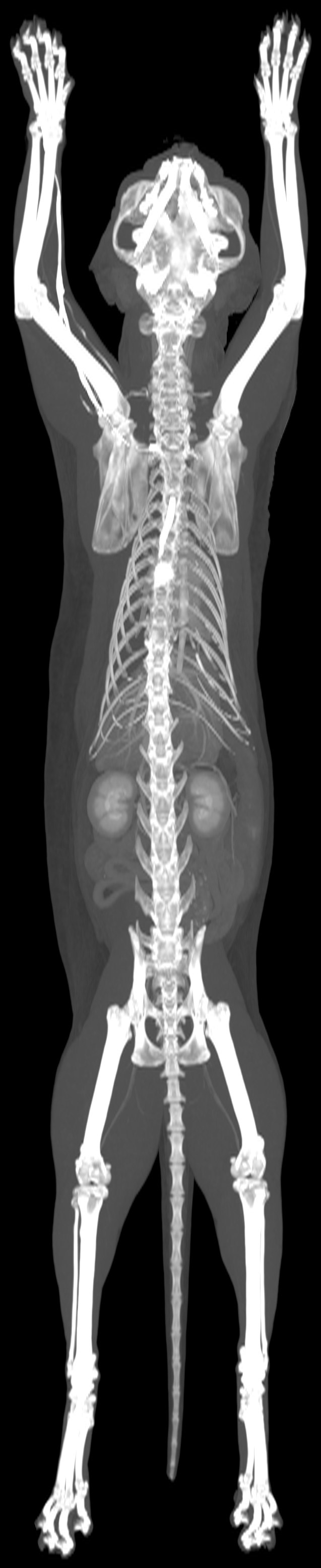
Maximum intensity projection of the whole scan

The whole scan in a single image: the skeleton, and the kidneys highlighted by the contrast agent.
Chest and lungs, Oncology

Breathing difficulties after tumour surgery, and what CT showed in the chest

Patient
cat, domestic shorthair, spayed female, 9 years
Examination
Whole-body CT, pre- and post-contrast
Date
May 2026
3D CT reconstruction

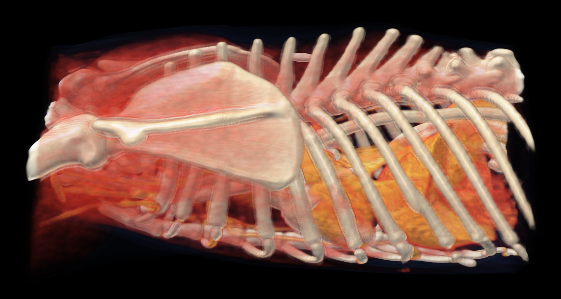
3D reconstruction of the chest, seen from the left: the ribs, the shoulder blade and, behind them, the heart highlighted by the contrast agent. Fluid in the chest does not show up in 3D; it can be seen on the cross-section below.

  • bone
  • strongest contrast uptake: blood vessels, heart, kidneys
  • organs and tissues with weaker contrast uptake, including tumours
  • skin and fat

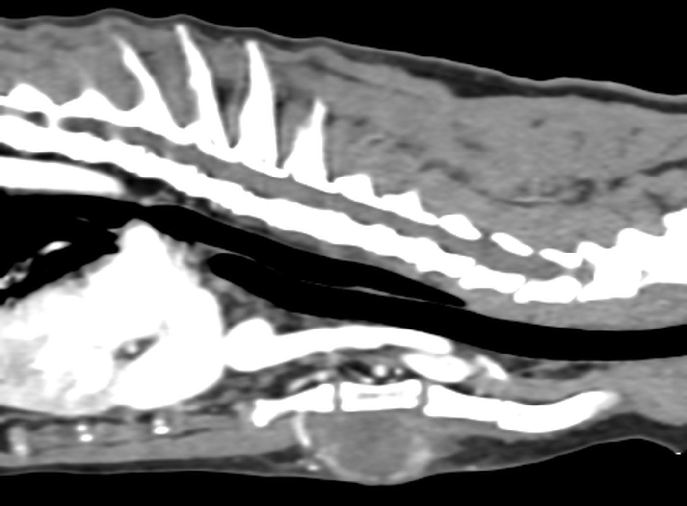
A nine-year-old cat had a lump removed from her thigh two months earlier; it turned out to be an adenocarcinoma. Later, because a diaphragmatic hernia was suspected, she underwent abdominal surgery. She then developed rapid abdominal breathing, her heart sounds were faint and her kidney values rose. Before the CT, about 100 ml of milky (chylous) fluid was drained from her chest.

The CT showed fluid in both halves of the chest and several lung lobes that were compressed and airless, most likely because of the pressure of the fluid itself. The lymph nodes in the chest were enlarged, as was one behind the knee. Next to the spine in the pelvic area, the CT found an irregularly thickened muscle, and both adrenal glands were also enlarged.

Transverse CT slice at the level of the finding

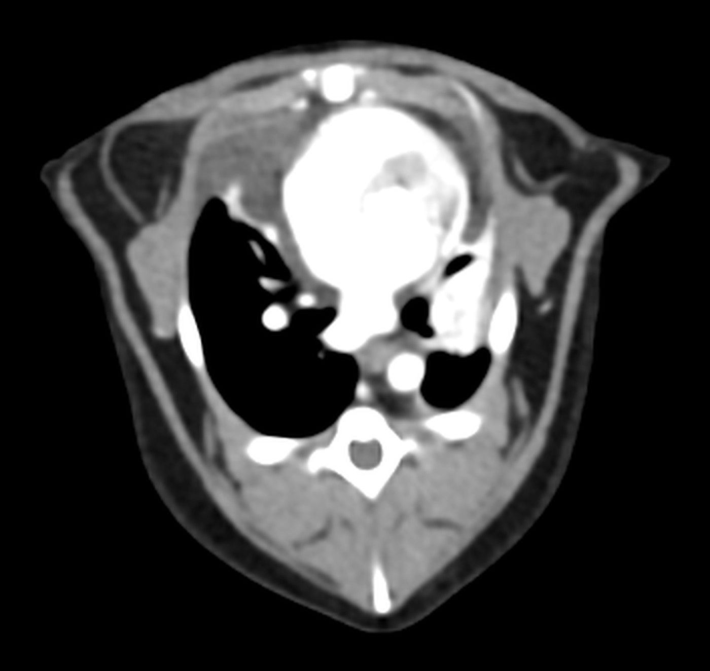
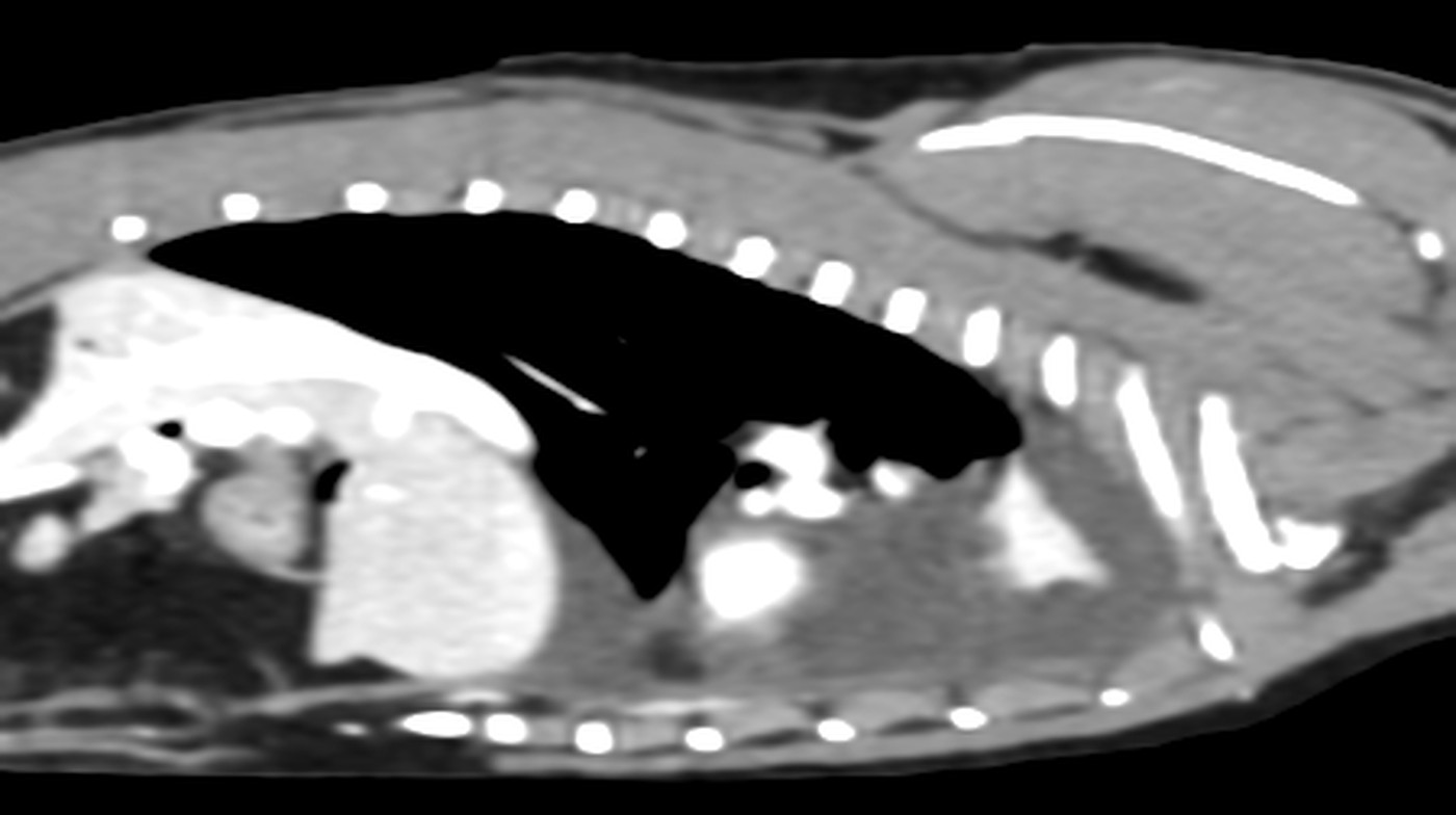
Cross-section through the chest: fluid around the lungs and compressed, airless lung lobes.

Longitudinal CT slice through the chest

Longitudinal slice through the chest: black is air in the lungs, and the grey fluid around them is compressing the lung lobes.

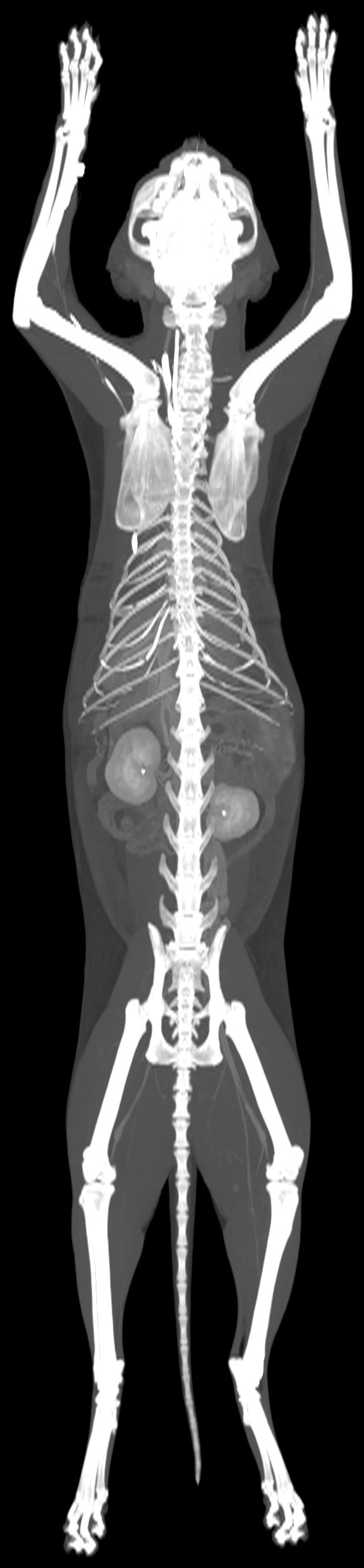
Maximum intensity projection of the whole scan

The whole scan in a single image: the skeleton, and the kidneys highlighted by the contrast agent.
Oncology, Chest and lungs, Abdomen

A lump on the chest. CT showed how deep it reached and also found something in the ureters

Patient
cat, Maine Coon, male, 10 years
Examination
CT of the thorax, abdomen and pelvis, pre- and post-contrast
Date
June 2026
3D CT reconstruction

3D reconstruction of the chest and abdomen, seen from the right. Low down, next to the breastbone, is the lump in the chest wall (orange-red); in the abdomen, the kidney and liver are strongly highlighted by the contrast agent (gold).

  • bone
  • strongest contrast uptake: blood vessels, heart, kidneys
  • organs and tissues with weaker contrast uptake, including tumours
  • skin and fat

A ten-year-old male cat had a lump next to his breastbone. The vet needed to know how big it was, what it involved and whether it had spread anywhere, and therefore recommended a CT scan of the chest, abdomen and pelvis.

The mass was about five centimetres in size. It lay under the skin next to the first three segments of the breastbone and was very probably already growing into the chest muscle. It did not extend into the chest cavity or reach the lungs. Alongside this, the CT picked up a small, irregular right kidney, a widened left ureter and signs that both ureters open in an unusual place, into the urethra instead of the bladder.

Transverse CT slice at the level of the finding

Cross-section through the chest: a mass under the skin next to the breastbone, reaching the chest muscle.

Longitudinal CT slice through the middle of the body

Longitudinal slice through the middle of the body: below the breastbone is a round lump in the chest wall.

Maximum intensity projection of the whole scan

The whole scan in a single image: the skeleton, and the kidneys highlighted by the contrast agent.
Oncology

A CT scan before treatment showed how far the sarcoma by the hip extended

Patient
dog, mixed breed, spayed female, 11 years
Examination
Whole-body CT, pre- and post-contrast
Date
August 2026
3D CT reconstruction

3D reconstruction of the pelvis and lower back, seen from the right: the lobulated tumour by the right hip (red), and the aorta and pelvic arteries (gold). Above the lumbar spine, a second mass, which lies on the left side, also shows through in a darker shade.

  • bone
  • strongest contrast uptake: blood vessels, heart, kidneys
  • organs and tissues with weaker contrast uptake, including tumours
  • skin and fat

An eleven-year-old female dog had a mass by her right hip, and a sample had confirmed it as a soft tissue sarcoma. Before a decision on treatment could be made, it was necessary to know how big it was, what it involved and whether it had spread anywhere. That is the purpose of a whole-body CT scan, known as oncological staging.

The tumour was about seven centimetres in size, was growing into the buttock muscles and extended into the pelvic cavity, where it was pressing slightly on the rectum. The bone beneath it was normal. In the lower back on the left, the CT found another mass, about seven centimetres in size. It found no distant metastases and no enlarged lymph nodes.

Transverse CT slice at the level of the finding

Cross-section through the pelvis: the lobulated tumour by the right hip, pushing the rectum aside.

Longitudinal CT slice through the pelvic region

Longitudinal slice through the tumour by the right hip: a lobulated mass that takes up the contrast agent unevenly.

Maximum intensity projection of the whole scan

The whole scan in a single image: the skeleton, and the kidneys highlighted by the contrast agent.
Oncology

Enlarged lymph nodes throughout the body. CT showed the extent of the disease and what needs to be ruled out

Patient
dog, Jack Russell Terrier, spayed female, 10 years
Examination
Whole-body CT, pre- and post-contrast
Date
May 2026
3D CT reconstruction

3D reconstruction of the trunk, seen from the left: the skeleton and, behind the ribs, the liver and the left kidney, which is highlighted by the contrast agent (gold).

  • bone
  • strongest contrast uptake: blood vessels, heart, kidneys
  • organs and tissues with weaker contrast uptake, including tumours
  • skin and fat

A ten-year-old female dog had had digestive problems for about a year. Then her lymph nodes became enlarged all over her body, she had a fever and a large mass grew under the skin on the right side of her chest. The vet recommended a whole-body CT scan to find out where in the body the disease was present.

The CT showed enlarged lymph nodes literally from head to hind legs: on the head and neck, in the chest, in the armpits, in the groin and behind the knees. Under the skin of the chest and abdomen it found flat masses on both sides; the largest, on the right flank, extended from the chest almost to the lower back. The liver, spleen and thymus were also enlarged.

Transverse CT slice at the level of the finding

Cross-section through the front part of the abdomen: an enlarged liver and thickened tissue under the skin along the back.

Longitudinal CT slice through the trunk

Longitudinal slice through the trunk, seen from above: under the skin on the side of the chest is a large, flat mass.

Maximum intensity projection of the whole scan

The whole scan in a single image: the skeleton, and the kidneys highlighted by the contrast agent.
Abdomen

A liver tumour was suspected. CT showed an enlarged liver with no distinct lesion

Patient
dog, mixed breed, neutered male, 13 years
Examination
Whole-body CT, pre- and post-contrast
Date
March 2026

A thirteen-year-old dog had raised liver values on blood tests, and a liver tumour was suspected. To find out whether there was a lesion in the liver and whether anything had spread in the body, the vet recommended a whole-body CT scan.

The liver was evenly and mildly enlarged, with no well-defined lesion, and the lymph nodes next to the liver were enlarged. The lungs were clear, and the CT found nothing significant in the other organs.

Ruling out disease, Abdomen

Low blood sugar and a suspected insulinoma. CT found no tumour, and the next step is a targeted one

Patient
female, 6 years
Examination
Whole-body CT, pre- and post-contrast
Date
September 2026

A six-year-old female patient was lethargic and had low blood sugar. The vet suspected an insulinoma, a tumour of the pancreas that releases too much insulin. She came for a CT scan to find out whether there was a visible lesion in the pancreas or elsewhere in the abdomen.

The CT found no lesion in the pancreas or in the other abdominal organs. Minor changes in the lungs may be related to the anaesthetic. An incidental finding was a mild narrowing, on the left, of the opening through which a nerve passes between the last lumbar vertebra and the sacrum.

Joints and bones

After a year of lameness in a front leg, the cause was found in the elbow

Patient
male, 5 years
Examination
CT of the forelimbs, pre- and post-contrast
Date
September 2026

For about a year, a five-year-old patient had repeatedly been lame on his left front leg. The elbow is difficult to assess on X-rays because the bones overlap there, so the vet recommended a CT scan of both front legs.

In the left elbow, the CT showed disease of a small bony projection of the ulna, called the medial coronoid process. This is a form of elbow dysplasia. The projection had not broken off, the joint surfaces fitted together correctly, and there was no new bone or soft tissue change around the joint.

Neurology, Head and neck

Seizures and walking in circles. CT found a lesion in the brain

Patient
dog, Staffordshire Bull Terrier, male, 11 years
Examination
CT of the head, pre- and post-contrast
Date
August 2026

For the past month, an eleven-year-old dog had been having epilepsy-like seizures and turning in circles to the right. The vet recommended a CT scan of the head to look for the cause in the brain and skull.

In the right hemisphere of the brain, the CT found a lesion about two centimetres in size that was pressing on its surroundings: the falx, the natural dividing wall between the hemispheres, was pushed slightly to the left. On the right, the thin bone that separates the brain from the nasal cavity was damaged, but the change had not grown into the nose. The CT also picked up breakdown of the necks of two teeth in the right upper jaw, with bone loss around one of them.

Oncology, Abdomen, Chest and lungs

Abdominal pain, weak hind legs and kidney problems shared a common cause

Patient
dog, mixed breed, male, 14 years
Examination
CT of the thorax, abdomen and pelvis, pre- and post-contrast
Date
February 2026

A fourteen-year-old dog had abdominal pain, was lethargic and weak in his hind legs, and had raised kidney values and enlarged lymph nodes. The vet recommended a CT scan of the chest, abdomen and pelvis to see how these problems were connected.

The CT showed enlarged lymph nodes in both the chest and the abdomen, an enlarged liver, both kidneys enlarged and interspersed with nodules, a thickened right ureter that was not draining the contrast agent, and tiny nodules in the lungs. It also found changes in the gallbladder, a little fluid in the abdomen with signs of inflammation of the abdominal lining, and an enlarged heart.

Oncology, Chest and lungs, Abdomen

Breathlessness and anaemia. CT showed the extent of the disease throughout the body

Patient
dog, German Shepherd Dog, spayed female, 11 years
Examination
CT of the neck, thorax, abdomen and pelvis, pre- and post-contrast
Date
August 2026

An eleven-year-old female German Shepherd came in with breathlessness, lethargy and anaemia. To find out what was behind this and how far the disease extended, the vet recommended a CT scan of her neck, chest, abdomen and pelvis.

The CT showed enlarged lymph nodes in several parts of the body, an enlarged liver and spleen, a small amount of fluid in the abdomen and changes in the lungs that were most consistent with pneumonia. Incidental findings were degenerative changes in the hip joints and the spine.