Nose and ear, Chest and lungs, Oncology
He came in because of his ear. CT also found a mass near the heart
- Patient
- dog, French Bulldog, male, 7 years
- Examination
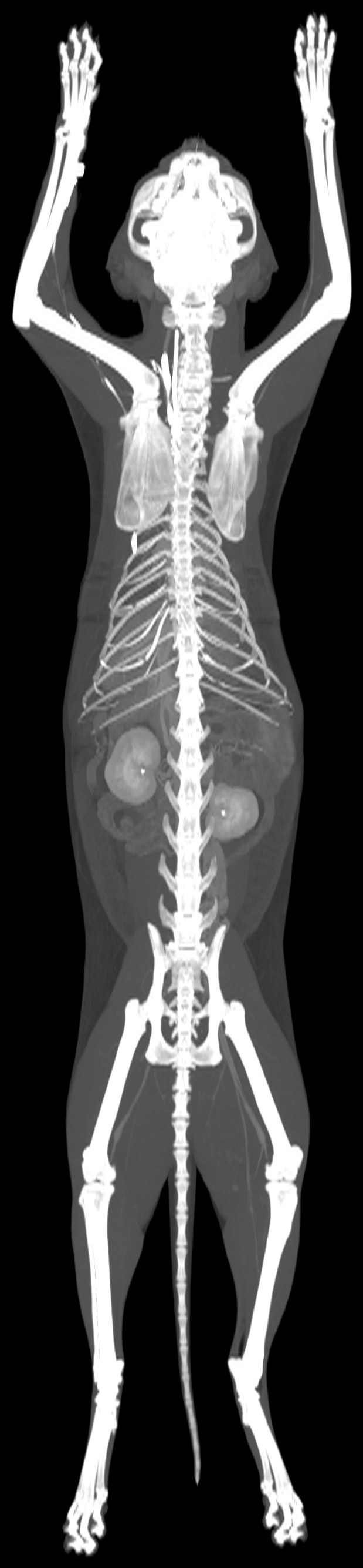
- Whole-body CT, pre- and post-contrast
- Date
- March 2026
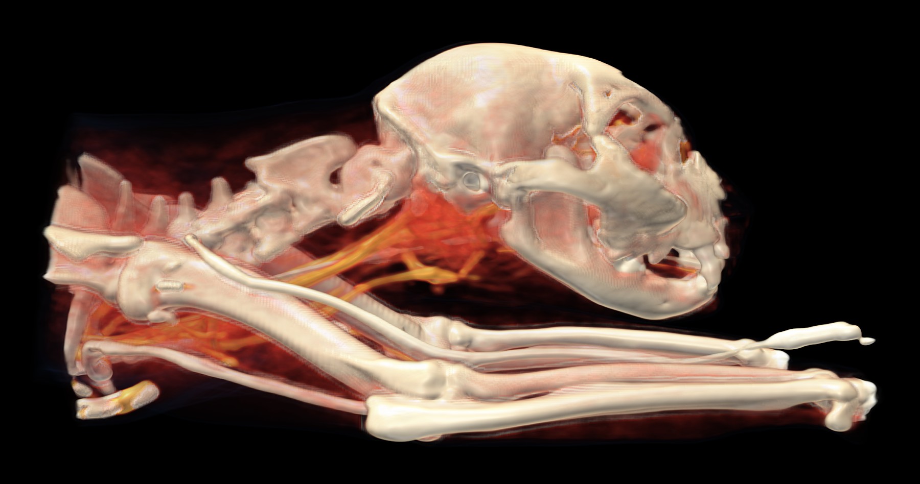
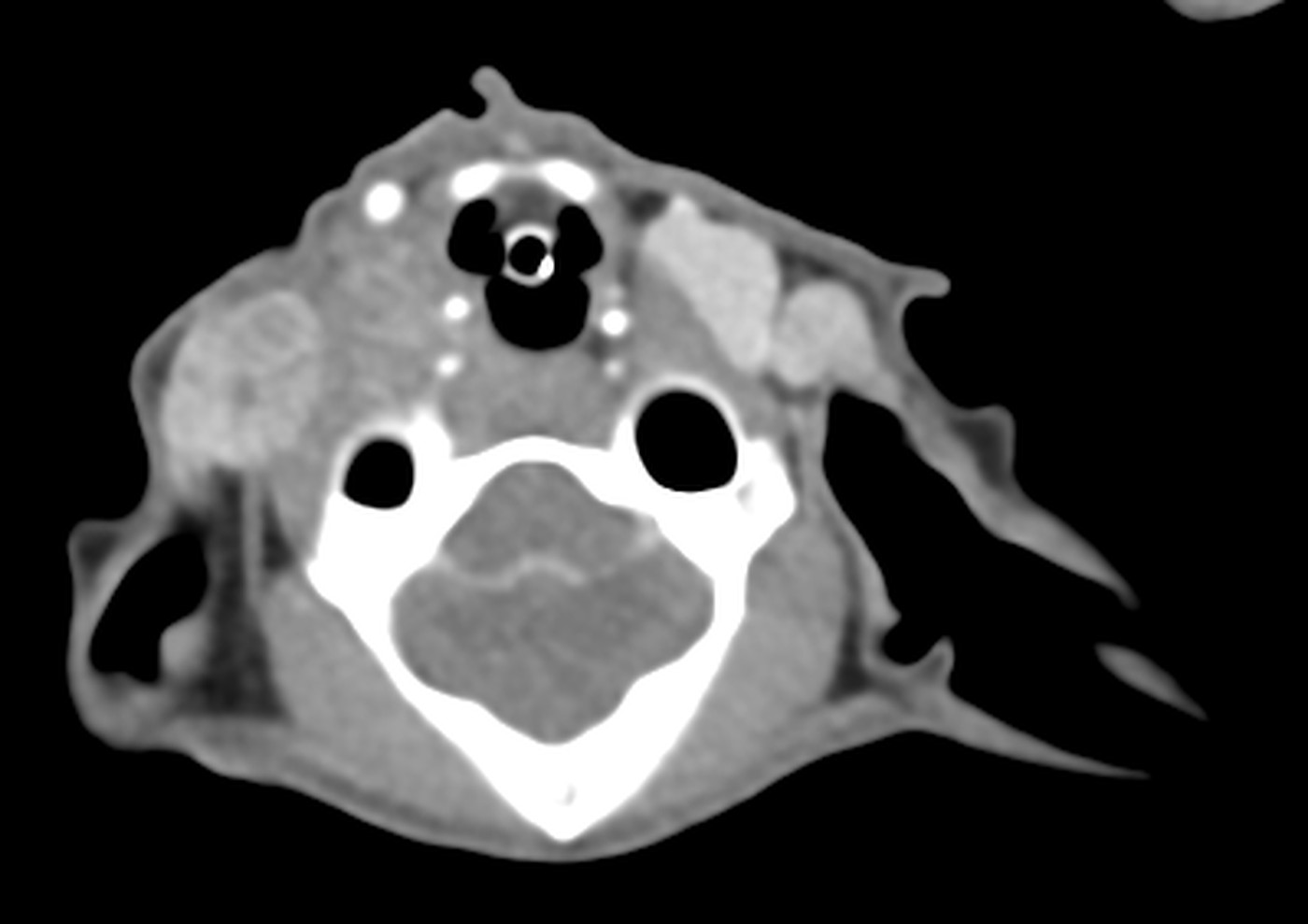
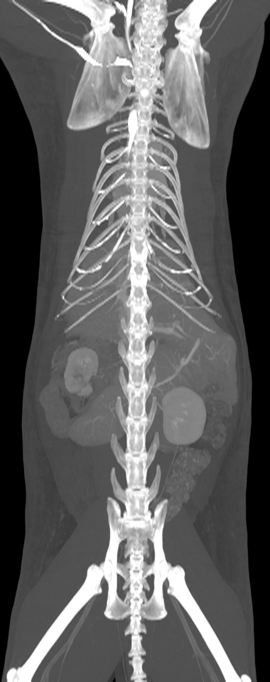
A seven-year-old French Bulldog had started tilting his head to the right and had a discharge from his right ear. To find out what was going on in the ear and deeper behind it, he had a whole-body CT scan.
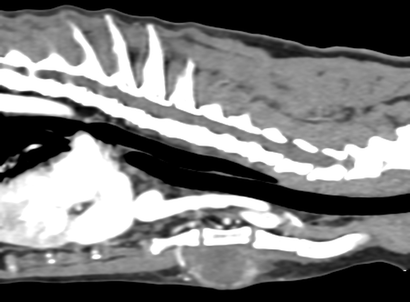
The CT confirmed inflammation of both outer ear canals, with a build-up of secretions on the right, and changes in the middle ear cavities that are common in flat-faced breeds. At the back of the nose it showed aberrant nasal turbinates, in other words tissue that should not normally be there.
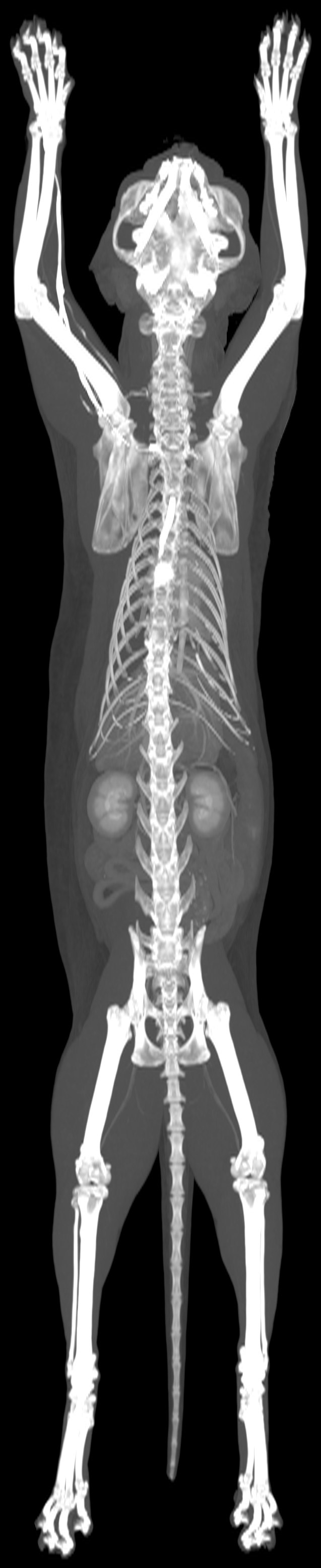
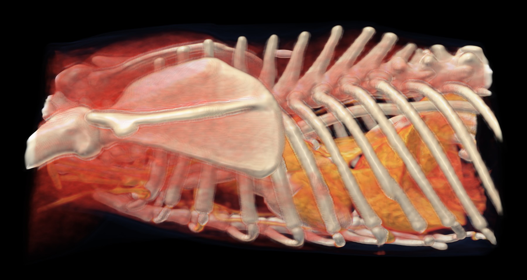
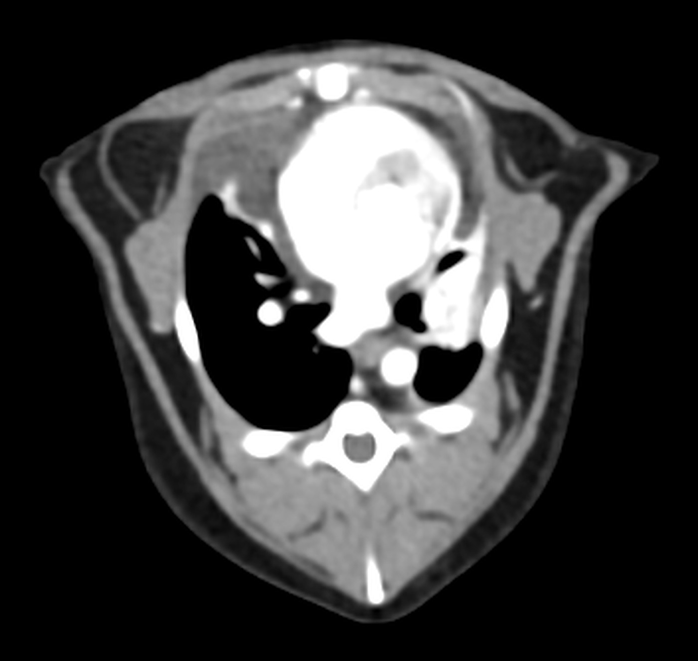
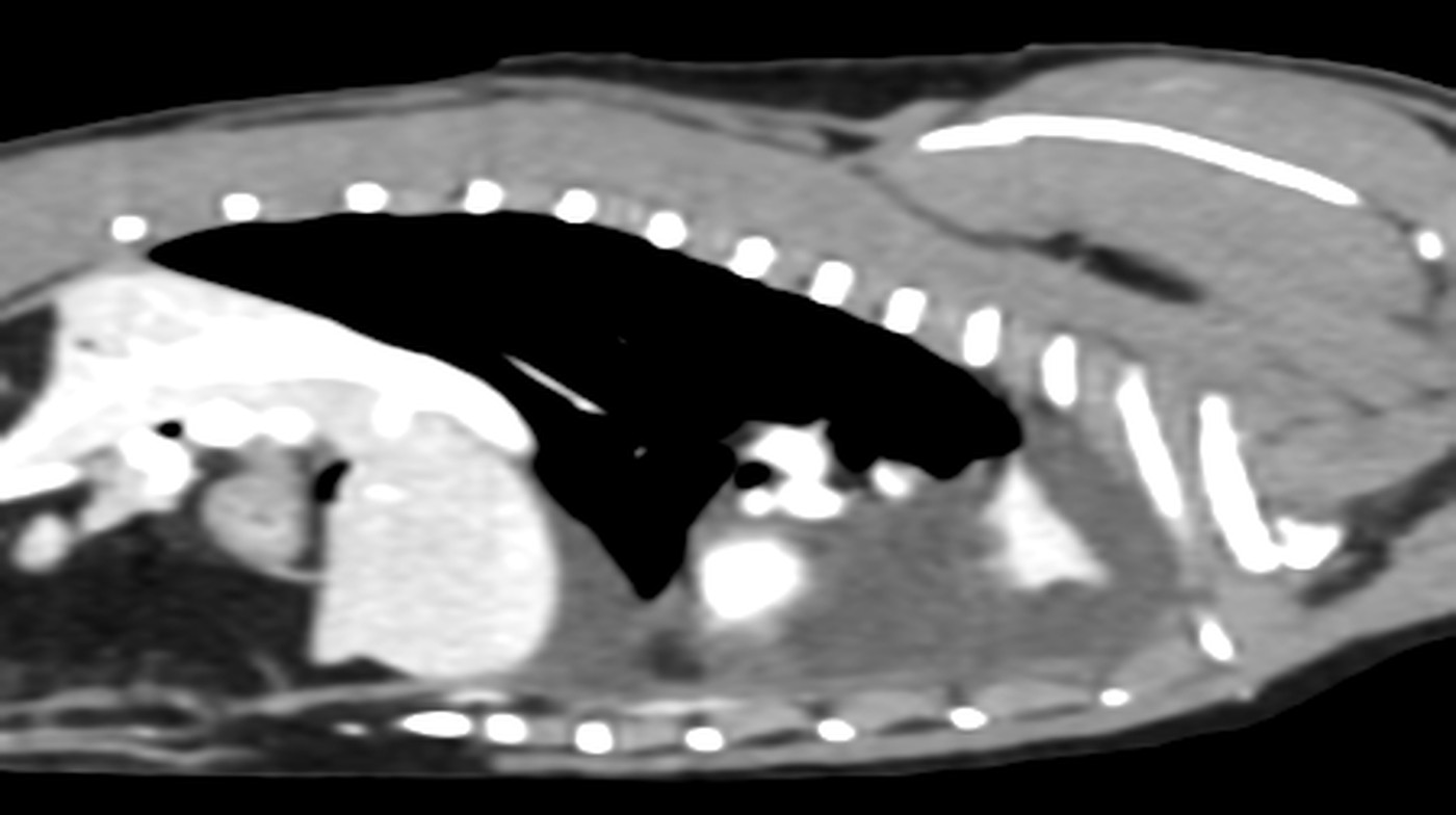
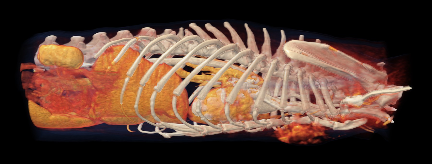
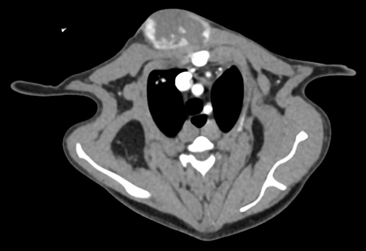
The most important finding, however, was one the dog had not come in for: a mass about five centimetres in size at the base of the heart, wrapping around the aorta and pressing on both pulmonary arteries, and next to it fluid in the sac around the heart. An examination focused only on the ear would have left it hidden.